
The Mechanisms of the 5 Senses and Sensory Disorders

Focus on the detailed mechanisms of vision, hearing, taste, smell, and touch. Study how sensory information is detected, transduced, and processed by the brain. Learn about common sensory disorders, their causes, symptoms, and treatments. Understanding these concepts is essential for grasping sensory perception and its effects on behavior and cognition. This comprehensive knowledge will help you excel in the AP Psychology exam
Learning Objectives
Understand the detailed processes by which the five senses—vision, hearing, taste, smell, and touch—detect and interpret sensory information. They should learn how each sensory system converts physical stimuli into neural signals and how these signals are processed in the brain. Additionally, students should recognize common sensory disorders, their causes, symptoms, and treatments. This knowledge is crucial for understanding sensory perception and its impact on behavior and cognition, essential for the AP Psychology exam
Vision
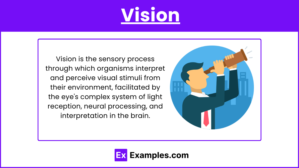
Mechanism of Vision
1. Light Detection
Cornea: Light enters the eye through the cornea, the eye's transparent, curved front surface that helps to focus incoming light.
Pupil: Light then passes through the pupil, the adjustable opening in the center of the iris. The size of the pupil is controlled by the iris, which adjusts to regulate the amount of light entering the eye.
Lens: Behind the pupil is the lens, a flexible structure that changes shape to focus light onto the retina. This process is called accommodation.
2. Photoreceptors
Retina: The retina, crucial for vision, contains rods and cones, photoreceptor cells essential for adaptation to varying light conditions.
Rods: Rods are responsible for vision in low light conditions (night vision) and peripheral vision. They do not detect color and are more numerous than cones.
Cones: Cones are responsible for color vision and function best in bright light. They are concentrated in the fovea, the central part of the retina.
3. Signal Processing
Phototransduction: Photoreceptors (rods and cones) convert light into electrical signals through a process called phototransduction. When light hits these cells, it triggers a chemical change in photopigments, leading to the generation of electrical impulses.
4. Transmission
Bipolar and Ganglion Cells: The electrical signals from the photoreceptors are first processed by bipolar cells and then by ganglion cells in the retina. Ganglion cells collect and transmit the visual information to the brain.
Optic Nerve: The axons of the ganglion cells form the optic nerve, which carries the visual information from the retina to the brain.
5. Brain Processing
Optic Chiasm: The optic nerves from both eyes meet at the optic chiasm, where the visual information is partially crossed to ensure that both hemispheres of the brain receive information from both eyes.
Thalamus: The visual signals are then relayed to the thalamus, specifically the lateral geniculate nucleus (LGN).
Visual Cortex: Finally, the signals are sent to the primary visual cortex (V1) in the occipital lobe of the brain. Here, the brain interprets the signals, enabling us to perceive shapes, colors, movement, and depth.
Sensory Disorders
1. Myopia (Nearsightedness)
Description: Difficulty seeing distant objects clearly because the eyeball is too long or the cornea is too curved, causing light to focus in front of the retina.
Correction: Glasses or contact lenses with concave lenses or refractive surgery.
2. Hyperopia (Farsightedness)
Description: Difficulty seeing close objects clearly because the eyeball is too short or the cornea is too flat, causing light to focus behind the retina.
Correction: Glasses or contact lenses with convex lenses alter light refraction, while refractive surgery reshapes molecules in the eye for vision correction.
3. Astigmatism
Description: Distorted or blurred vision at all distances due to an irregularly shaped cornea or lens.
Correction: Glasses or contact lenses with a special cylindrical lens or refractive surgery.
4. Cataracts
Description: Clouding of the lens, leading to blurred or dim vision.
Causes: Aging, injury, prolonged exposure to UV light, or certain medical conditions.
Treatment: Surgical removal of the cloudy lens and replacement with an artificial lens.
5. Glaucoma
Description: Damage to the optic nerve, often due to increased intraocular pressure, leading to vision loss.
Types: Open-angle glaucoma (chronic) and angle-closure glaucoma (acute).
Treatment: Medications, laser treatment, or surgery to lower intraocular pressure.
6. Macular Degeneration
Description: Damage to the macula, affected by saturated fats, results in central vision loss due to retinal impairment.
Types: Dry (atrophic) and wet (neovascular).
Treatment: Vitamins for dry macular degeneration; laser therapy or injections for wet macular degeneration.
7. Retinal Detachment
Description: The retina separates from the back of the eye, leading to potential vision loss.
Causes: Trauma, advanced diabetes, or inflammatory disorders.
Treatment: Immediate surgical intervention to reattach the retina.
Hearing
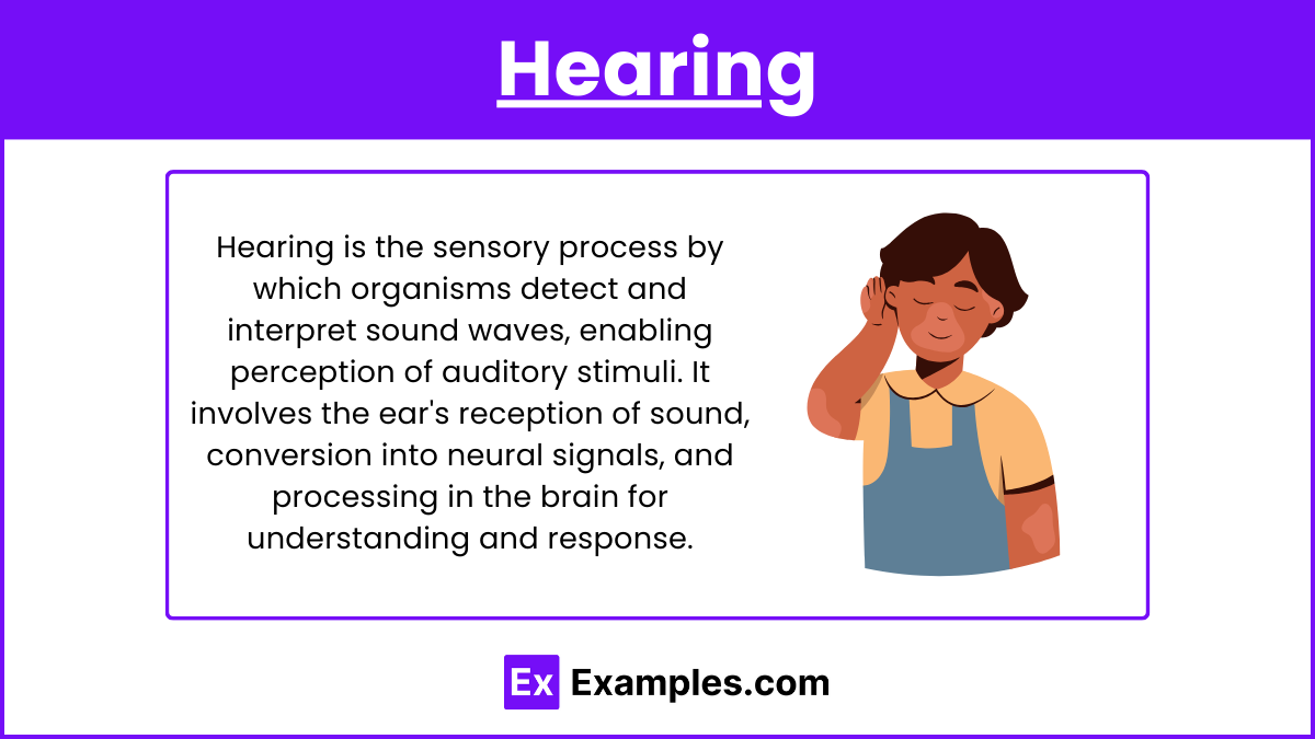
Mechanism of Hearing
1. Sound Wave Detection
Outer Ear: Sound waves are collected by the outer ear, also known as the pinna or auricle, and travel through the ear canal to the eardrum.
Eardrum (Tympanic Membrane): The eardrum vibrates in response to the sound waves, converting them into mechanical vibrations.
2. Middle Ear
Ossicles: The middle ear contains three small bones called the ossicles (malleus, incus, and stapes). These bones amplify the vibrations from the eardrum and transmit them to the inner ear.
Eustachian Tube: This tube connects the middle ear to the throat, helping to equalize pressure on both sides of the eardrum.
3. Inner Ear
Cochlea: The stapes vibrates against the oval window of the cochlea, a fluid-filled, snail-shaped structure in the inner ear. The vibrations create waves in the cochlear fluid.
Hair Cells: Inside the cochlea, hair cells (sensory receptors) are stimulated by the fluid waves. These hair cells convert the mechanical vibrations into electrical signals.
4. Signal Processing
Organ of Corti: Located within the cochlea, the Organ of Corti contains the hair cells and converts the mechanical vibrations into neural signals.
Basilar Membrane: Different frequencies of sound waves stimulate different parts of the basilar membrane, allowing for the perception of pitch.
5. Transmission
Auditory Nerve: The electrical signals generated by the hair cells are transmitted to the brain via the auditory nerve (also known as the cochlear nerve).
Brain Processing: The signals travel through the brainstem to the thalamus and then to the auditory cortex in the temporal lobe, where they are interpreted as sound.
Sensory Disorders
1. Conductive Hearing Loss
Description: Caused by problems in the outer or middle ear that prevent sound from being conducted to the inner ear.
Causes: Ear infections, fluid in the middle ear, perforated eardrum, or blockage by earwax.
Treatment: Medical treatment, surgery, or hearing aids.
2. Sensorineural Hearing Loss
Description: Caused by damage to the inner ear (cochlea) or auditory nerve.
Causes: Aging, exposure to loud noises, genetic factors, or illnesses.
Treatment: Hearing aids, cochlear implants, or other assistive devices.
3. Mixed Hearing Loss
Description: A combination of conductive and sensorineural hearing loss.
Causes: A mix of issues affecting both the outer/middle ear and the inner ear/auditory nerve.
Treatment: A combination of medical treatments, hearing aids, or surgery.
4. Tinnitus
Description: Perception of noise or ringing in the ears without an external sound source.
Causes: Exposure to loud noise, ear infections, earwax buildup, or other medical conditions.
Treatment: Treating underlying conditions, sound therapy, or medication.
5. Meniere's Disease
Description: Inner ear disorder causing episodes of vertigo, tinnitus, hearing loss, and a feeling of fullness in the ear.
Causes: Abnormal fluid buildup in the inner ear.
Treatment: Medications to reduce symptoms, dietary changes, or in severe cases, surgery.
Taste
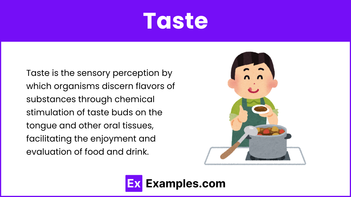
Mechanism of Taste
1. Taste Buds
Location: Taste buds are located on the papillae of the tongue, as well as on the roof of the mouth, the inner cheeks, and the throat.
Types of Papillae: There are four types of papillae that contain taste buds: fungiform, foliate, circumvallate, and filiform (though filiform papillae do not contain taste buds).
2. Taste Receptors
Function: Each taste bud contains 50-100 taste receptor cells that are responsible for detecting different taste modalities.
Five Basic Tastes: The five primary tastes are sweet, sour, salty, bitter, and umami (savory).
3. Signal Processing
Chemical Detection: When food or drink enters the mouth, it dissolves in saliva and comes into contact with taste receptor cells. These cells detect the chemical compounds responsible for different tastes.
Transduction: The taste receptor cells convert these chemical signals into electrical impulses through a process known as transduction.
4. Transmission
Cranial Nerves: The electrical signals are transmitted to the brain via three cranial nerves:
Facial Nerve (VII): Carries taste information from the front two-thirds of the tongue.
Glossopharyngeal Nerve (IX): Carries taste information from the back one-third of the tongue.
Vagus Nerve (X): Carries taste information from the throat and epiglottis.
5. Brain Processing
Medulla Oblongata: The taste signals are first processed in the medulla oblongata.
Thalamus: Signals are then relayed to the thalamus.
Gustatory Cortex: Finally, the signals reach the gustatory cortex in the frontal lobe, where they are interpreted as distinct tastes.
Sensory Disorders
1. Ageusia
Description: Complete loss of taste function.
Causes: Infections, head injuries, neurological disorders, or certain medications.
Treatment: Addressing underlying causes or discontinuing medications causing the condition.
2. Hypogeusia
Description: Reduced ability to taste.
Causes: Aging, zinc deficiency, medications, or nerve damage.
Treatment: Nutritional supplements, changing medications, or treating underlying conditions.
3. Dysgeusia
Description: Distorted taste perception, often resulting in a metallic, bitter, or salty taste.
Causes: Infections, chemotherapy, medications, or oral health issues.
Treatment: Managing the underlying cause, improving oral hygiene, or adjusting medications.
4. Burning Mouth Syndrome
Description: Persistent burning sensation in the mouth without an obvious cause.
Causes: Nutritional deficiencies, hormonal changes, nerve damage, or psychological factors.
Treatment: Nutritional supplements, medications for nerve pain, or therapy for psychological factors.
Smell
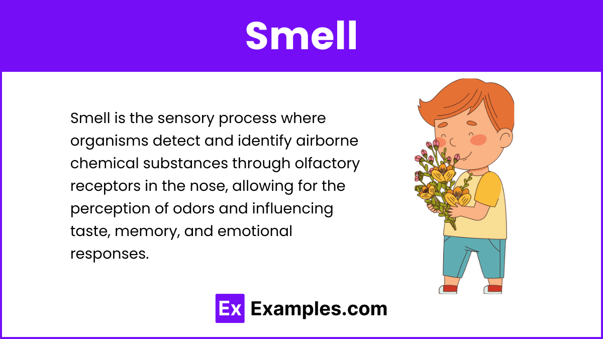
Mechanism
Olfactory Receptors: Located in the nasal cavity, these receptors detect airborne chemicals.
Signal Processing: Olfactory receptors convert chemical stimuli into electrical signals.
Transmission: Signals are transmitted to the olfactory bulb and then to the brain.
Brain Processing: The olfactory cortex and limbic system process these signals, linking smell to memory and emotion.
Sensory Disorders
Anosmia: Loss of the sense of smell.
Hyposmia: Reduced ability to smell.
Parosmia: Distorted smell perception.
Phantosmia: Perception of smells that aren't present.
Touch
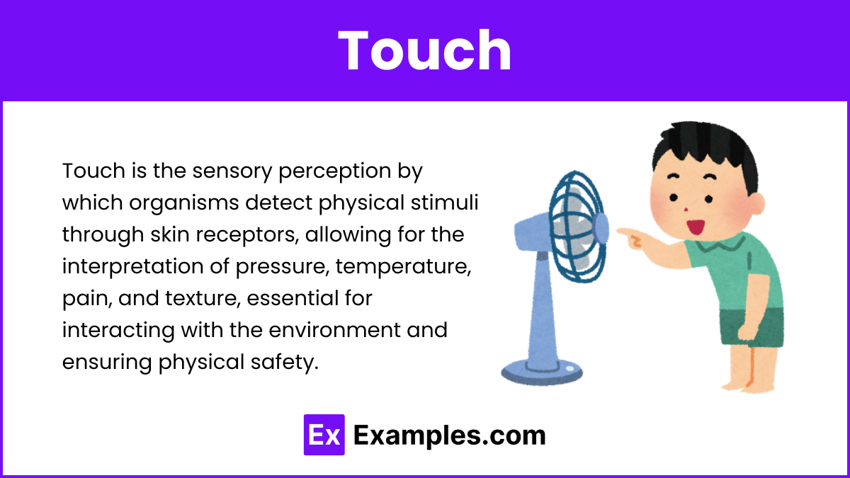
Mechanism of Touch
1. Receptors in the Skin
Types of Receptors: The skin contains various types of sensory receptors that detect different stimuli:
Mechanoreceptors: Respond to mechanical pressure or distortion.
Merkel Cells: Detect light touch and texture.
Meissner's Corpuscles: Detect light touch and vibrations.
Ruffini Endings: Detect skin stretch and sustained pressure.
Pacinian Corpuscles: Detect deep pressure and high-frequency vibrations.
Thermoreceptors: Respond to changes in temperature.
Krause End Bulbs: Detect cold temperatures.
Ruffini Endings: Also respond to heat.
Nociceptors: Detect pain from physical damage or inflammation.
2. Signal Processing
Transduction: When receptors are stimulated by touch, temperature, or pain, they convert these stimuli into electrical signals through transduction.
Peripheral Nerves: These electrical signals are transmitted from the skin to the spinal cord via peripheral nerves.
3. Transmission
Afferent Pathways: The signals travel through afferent nerve fibers (sensory neurons) to the spinal cord.
Spinal Cord: In the spinal cord, signals are processed and relayed to the brain through ascending pathways such as the spinothalamic tract (for pain and temperature) and the dorsal column-medial lemniscus pathway (for touch and proprioception).
4. Brain Processing
Thalamus: The signals reach the thalamus, which acts as a relay station, directing them to appropriate areas of the brain.
Somatosensory Cortex: Located in the parietal lobe, the primary somatosensory cortex interprets the signals, allowing us to perceive touch, temperature, and pain. This area is organized somatotopically, meaning different parts of the cortex correspond to sensations from different parts of the body.
Sensory Disorders
1. Neuropathy
Description: Damage to peripheral nerves causing numbness, tingling, or pain.
Causes: Diabetes, infections, autoimmune diseases, or physical trauma.
Treatment: Managing underlying conditions, medications for pain relief, or physical therapy.
2. Hyperesthesia
Description: Increased sensitivity to sensory stimuli.
Causes: Neurological disorders, infections, or certain medications.
Treatment: Treating the underlying cause, pain management, or sensory integration therapy.
3. Hypoesthesia
Description: Reduced sensitivity to sensory stimuli.
Causes: Nerve damage, stroke, or certain medical conditions.
Treatment: Addressing underlying causes, occupational therapy, or sensory re-education.
4. Tactile Agnosia (Astereognosis)
Description: Inability to recognize objects by touch despite normal tactile sensation.
Causes: Brain damage, particularly in the parietal lobe, often due to stroke or trauma.
Treatment: Occupational therapy to improve tactile recognition skills.
5. Allodynia
Description: Pain from stimuli that do not usually provoke pain, such as light touch.
Causes: Neuropathic pain conditions, fibromyalgia, or after nerve injury.
Treatment: Medications for pain relief, physical therapy, or desensitization techniques.