

The Study of the Brain and Research Techniques for Studying its Structure and Function

Understanding the brain's structure and function is crucial in AP Psychology. The brain, composed of various interconnected regions, governs behavior, emotions, and cognitive processes. Research techniques like MRI, PET scans, and EEG are vital for studying these aspects. These methods allow psychologists to visualize brain activity, identify structural abnormalities, and understand neural mechanisms. Mastering these concepts and techniques is essential for success in the AP Psychology exam, providing a foundation for exploring complex psychological phenomena.
Learning Objectives
Gain a comprehensive understanding of the brain's structure and function, including key areas such as the cerebrum, cerebellum, and brainstem. Study neural firing mechanisms, including how neurons communicate through electrical impulses and neurotransmitters. Explore the influence of medication on brain activity, including how different drugs can alter neural processes and behavior. Learn and apply key research techniques used to study the brain, such as MRI (Magnetic Resonance Imaging), PET (Positron Emission Tomography) scans, and EEG (Electroencephalography). These objectives are essential for excelling in the AP Psychology exam.
Key Brain Structures
Understanding the key structures of the brain and their functions is essential for AP Psychology. Here, we break down the main components and their roles.
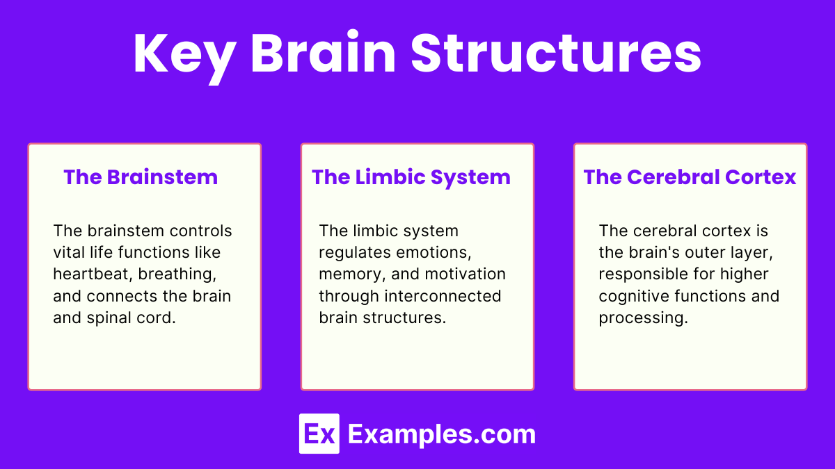
The Brainstem
The brainstem is responsible for basic life functions and acts as a relay center connecting the cerebrum and cerebellum to the spinal cord. It consists of the medulla oblongata, pons, and midbrain.
Medulla Oblongata
Function: Controls vital autonomic functions such as heartbeat, breathing, and blood pressure.
Location: Base of the brainstem, just above the spinal cord.
Pons
Function: Assists in regulating sleep, respiration, swallowing, bladder control, hearing, equilibrium, taste, eye movement, facial expressions, and posture.
Location: Above the medulla, connects the medulla to the midbrain.
Midbrain
Function: Involved in motor movement, particularly movements of the eye, and in auditory and visual processing.
Location: Above the pons, part of the brainstem.
The Limbic System
The limbic system is a complex set of structures in the brain that lies on both sides of the thalamus, just under the cerebrum. It is involved in many of our emotions and motivations, particularly those related to survival such as fear and anger. It also plays a key role in the formation of memories. Here, we explore the primary components of the limbic system and their functions.
Key Components of the Limbic System
Amygdala
Function: The amygdala is critical for emotion processing. It is particularly involved in the detection of fear and the formation of fear-related memories. The amygdala also plays a role in the recognition of emotional expressions in others.
Location: Deep within the temporal lobes, one in each hemisphere.
Hippocampus
Function: The hippocampus is essential for the formation of new long-term memories and is involved in spatial navigation. It helps convert short-term memories into long-term ones, a process crucial for learning.
Location: Located in the medial temporal lobe, extending from the amygdala.
Hypothalamus
Function: The hypothalamus regulates many bodily functions, including temperature, hunger, thirst, and circadian rhythms. It also controls the autonomic nervous system and the endocrine system through its connection to the pituitary gland.
Location: Located below the thalamus and above the brainstem.
Thalamus
Function: The thalamus acts as the brain's relay station, channeling sensory and motor signals to the cerebral cortex. It also regulates consciousness, sleep, and alertness.
Location: Situated at the top of the brainstem, near the center of the brain.
Cingulate Gyrus
Function: The cingulate gyrus is involved in processing emotions and behavior regulation. It also helps regulate autonomic motor function.
Location: Located above the corpus callosum.
Fornix
Function: The fornix acts as a major output tract of the hippocampus. It is involved in the transfer of information within the limbic system, particularly from the hippocampus to the mammillary bodies and septal nuclei.
Location: An arching, fibrous band extending from the hippocampus to the hypothalamus.
Mammillary Bodies
Function: The mammillary bodies play a role in recollective memory. They are involved in the processing of recognition memory and spatial memory.
Location: Located at the ends of the anterior arches of the fornix.
Functions of the Limbic System
Emotion Processing : The limbic system is heavily involved in emotional processing. The amygdala, for example, is essential for recognizing emotional stimuli and triggering appropriate responses. Damage to the amygdala can result in a lack of fear or inappropriate emotional responses.
Memory Formation : The hippocampus is crucial for the formation and retrieval of memories. It helps convert short-term memories into long-term ones, and its dysfunction can lead to difficulties in forming new memories (anterograde amnesia).
Autonomic and Endocrine Regulation : The hypothalamus controls various autonomic processes, such as hunger, thirst, and body temperature, and it links the nervous system to the endocrine system via the pituitary gland. This regulation is vital for maintaining homeostasis.
Motivational Behaviors : The limbic system also influences motivational behaviors related to survival, such as eating, drinking, and reproductive behaviors. It helps ensure that these behaviors are performed in response to internal needs and external stimuli.
The Cerebral Cortex
The cerebral cortex is the outermost layer of the brain, characterized by its wrinkled appearance, which increases surface area and allows for greater cognitive capacity. It is responsible for many of the brain’s higher-order functions, including thought, perception, and voluntary movement. The cortex is divided into two hemispheres, each containing four lobes with distinct functions.
Four Lobes of the Cerebral Cortex
1. Frontal Lobes
Function: The frontal lobes are involved in a variety of complex behaviors, including planning, decision-making, problem-solving, and controlling behavior and emotions. They also play a crucial role in speech production and voluntary movement.
Key Areas:
Prefrontal Cortex: Associated with planning, reasoning, and social behavior.
Motor Cortex: Controls voluntary movements.
Broca’s Area: Involved in speech production (usually located in the left hemisphere).
Location: Located at the front of the brain, just behind the forehead.
2. Parietal Lobes
Function: The parietal lobes process sensory information from the body, such as touch, temperature, and pain. They are also involved in spatial orientation and the coordination of movement.
Key Areas:
Somatosensory Cortex: Processes tactile information from the body.
Posterior Parietal Cortex: Involved in spatial reasoning and attention.
Location: Located near the top and back of the brain, behind the frontal lobes.
3. Occipital Lobes
Function: The occipital lobes are primarily responsible for visual processing. They receive and interpret input from the eyes, enabling us to understand and respond to visual information.
Key Areas:
Primary Visual Cortex: The main area for receiving visual information.
Visual Association Areas: Involved in interpreting and recognizing visual stimuli.
Location: Located at the back of the brain.
4. Temporal Lobes
Function: The temporal lobes are involved in processing auditory information and are crucial for understanding language. They also play a significant role in memory formation and retrieval.
Key Areas:
Auditory Cortex: Processes sounds and is involved in hearing.
Wernicke’s Area: Important for language comprehension (usually located in the left hemisphere).
Hippocampus: Embedded within the temporal lobe, crucial for memory formation.
Location: Located on the sides of the brain, near the temples.
Functions of the Cerebral Cortex
Sensory Processing : The cerebral cortex is responsible for processing sensory information from various parts of the body. The somatosensory cortex in the parietal lobe, for example, processes touch, pain, and temperature.
Motor Control : The motor cortex, located in the frontal lobe, controls voluntary movements. Specific areas of the motor cortex correspond to different parts of the body, allowing for precise and coordinated movements.
Language and Communication : The cerebral cortex includes areas critical for language production and comprehension. Broca’s area (in the frontal lobe) is involved in speech production, while Wernicke’s area (in the temporal lobe) is essential for understanding spoken and written language.
Higher-Order Functions : The prefrontal cortex in the frontal lobes is involved in complex cognitive functions such as planning, decision-making, and social behavior. These higher-order functions distinguish humans from other animals and are crucial for adaptive and goal-directed behavior.
Memory : The hippocampus, part of the temporal lobes, plays a key role in forming and retrieving memories. It is essential for converting short-term memories into long-term ones, allowing us to retain and recall past experiences.
Research Techniques
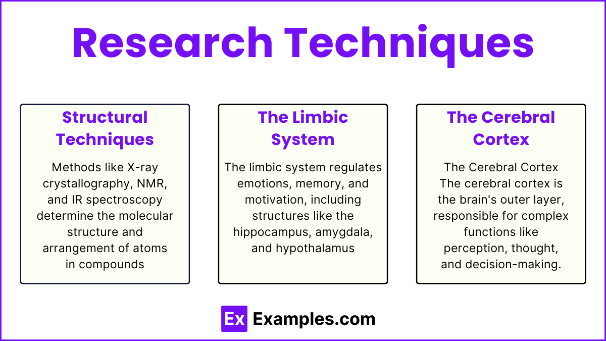
Structural Techniques
1. Magnetic Resonance Imaging (MRI)
Principle: MRI uses strong magnetic fields and radio waves to generate detailed images of the brain's internal structures. The technique exploits the magnetic properties of hydrogen atoms in the body.
Process:
The patient lies inside an MRI scanner, which creates a strong magnetic field.
Radio waves are sent through the body, causing hydrogen atoms to emit signals.
The scanner detects these signals and a computer processes them to create detailed images.
Advantages:
High-resolution images.
Non-invasive.
No exposure to ionizing radiation.
Disadvantages:
Expensive.
Not suitable for patients with metal implants or certain medical devices.
Can be uncomfortable for claustrophobic individuals.
2. Computerized Tomography (CT) Scan
Principle: CT scans use X-rays to create cross-sectional images of the brain. Multiple X-ray measurements are taken from different angles and processed by a computer to produce detailed images.
Process:
The patient lies on a table that slides into a CT scanner.
The scanner rotates around the head, taking multiple X-ray images.
A computer combines these images to create cross-sectional slices of the brain.
Advantages:
Quick and widely available.
Effective for detecting brain injuries, tumors, and structural abnormalities.
Disadvantages:
Exposure to ionizing radiation.
Lower resolution compared to MRI.
Limited ability to differentiate between similar types of soft tissue.
3. Diffusion Tensor Imaging (DTI)
Principle: DTI is a type of MRI that measures the diffusion of water molecules in brain tissue. It provides detailed images of the brain's white matter tracts, which are crucial for understanding connectivity and communication between different brain regions.
Process:
The patient undergoes an MRI scan with specific sequences that measure water diffusion.
Data are processed to create images showing the orientation and integrity of white matter tracts.
Advantages:
High-resolution images of white matter pathways.
Useful for studying brain connectivity and diagnosing conditions like multiple sclerosis.
Non-invasive and no radiation exposure.
Disadvantages:
Complex data analysis required.
Expensive and time-consuming.
Functional Techniques
Principle: fMRI measures brain activity by detecting changes in blood flow. When a brain region is more active, it consumes more oxygen, and blood flow to that area increases. fMRI detects these changes using the Blood Oxygen Level Dependent (BOLD) signal.
Process:
The patient lies inside an MRI scanner.
The scanner captures images of brain activity by measuring blood flow changes.
A computer processes these images to create maps of brain activity.
Advantages:
Non-invasive with no radiation exposure.
High spatial resolution.
Can map brain activity in response to specific tasks.
Disadvantages:
Expensive.
Requires the patient to remain still.
Temporal resolution is lower compared to EEG and MEG.
2. Positron Emission Tomography (PET) Scan
Principle: PET scans measure brain activity by detecting radioactive tracers injected into the bloodstream. These tracers accumulate in areas of high brain activity, allowing researchers to visualize metabolic processes.
Process:
A radioactive tracer is injected into the patient’s bloodstream.
The patient lies in a PET scanner.
The scanner detects the gamma rays emitted by the tracer and constructs images showing areas of high activity.
Advantages:
Can measure metabolic activity and neurotransmitter function.
Useful for diagnosing brain disorders and cancers.
Disadvantages:
Exposure to radiation.
Lower spatial and temporal resolution compared to fMRI.
Expensive and not as widely available.
3. Electroencephalogram (EEG)
Principle: EEG measures electrical activity in the brain through electrodes placed on the scalp. It records the brain's electrical signals, which are generated by neurons firing.
Process:
Electrodes are placed on the patient’s scalp.
The electrodes detect electrical signals and transmit them to a computer.
The computer records and displays the signals as waveforms.
Advantages:
Non-invasive and relatively inexpensive.
Excellent temporal resolution, capturing brain activity in real-time.
Useful for diagnosing epilepsy, sleep disorders, and brain function.
Disadvantages:
Poor spatial resolution.
Primarily measures surface activity, not deep brain structures.
4. Magnetoencephalography (MEG)
Principle: MEG measures the magnetic fields produced by neuronal activity in the brain. It detects these fields using highly sensitive devices called SQUIDs (Superconducting Quantum Interference Devices).
Process:
The patient sits or lies under an MEG scanner.
The scanner detects magnetic fields generated by brain activity.
A computer processes these signals to create maps of brain activity.
Advantages:
Non-invasive with excellent temporal resolution.
High spatial resolution, better than EEG.
Can pinpoint activity in specific brain areas.
Disadvantages:
Very expensive and not widely available.
Requires a magnetically shielded room.
Invasive Techniques
1. Lesion Studies
Principle: Lesion studies involve intentionally damaging or observing naturally occurring damage to specific brain areas to study the effects on behavior and cognitive function.
Process:
Lesions can be created surgically, chemically, or electrically in animal models.
Alternatively, researchers study patients with naturally occurring brain damage from injury, stroke, or disease.
Behavioral and cognitive assessments are conducted to understand the function of the damaged area.
Advantages:
Provides direct evidence of brain region function.
Helps identify brain areas critical for specific behaviors and cognitive processes.
Disadvantages:
Ethical concerns in humans.
Damage is often irreversible.
Results can be confounded by the brain's ability to compensate for damage (neuroplasticity).
2. Electrical Stimulation
Principle: Electrical stimulation involves applying small electrical currents to specific brain areas to observe the effects on behavior and cognitive function.
Process:
Electrodes are surgically implanted in targeted brain regions.
Electrical currents are delivered to stimulate neurons.
Researchers observe changes in behavior, perception, or cognitive function.
Advantages:
Can precisely target specific brain areas.
Reversible effects, as stimulation can be turned on and off.
Used therapeutically in conditions like Parkinson's disease (deep brain stimulation).
Disadvantages:
Surgical risks, including infection and bleeding.
Ethical concerns.
Potential for unintended effects on surrounding brain tissue.
3. Deep Brain Stimulation (DBS)
Principle: DBS involves implanting electrodes in specific brain regions to modulate neural activity and treat neurological and psychiatric conditions.
Process:
Electrodes are surgically implanted in targeted brain areas, such as the basal ganglia.
Electrodes are connected to a pulse generator implanted under the skin.
Electrical impulses are delivered to modulate neural activity.
Advantages:
Effective treatment for conditions like Parkinson's disease, essential tremor, and dystonia.
Adjustable and reversible stimulation.
Can significantly improve quality of life for patients with movement disorders.
Disadvantages:
Invasive surgery with associated risks.
High cost and potential for device complications.
Requires regular follow-up and device maintenance.
4. Intracranial Electroencephalography (iEEG)
Principle: iEEG involves placing electrodes directly on the brain's surface or within the brain to record electrical activity with high precision.
Process:
Electrodes are surgically implanted on the brain's surface (subdural) or within the brain tissue (depth electrodes).
Electrical activity is recorded during various tasks or states of rest.
Data is analyzed to identify patterns of neural activity and localize functions or abnormalities.
Advantages:
High spatial and temporal resolution.
Direct recording from brain tissue, reducing signal interference from the skull and scalp.
Essential for pre-surgical planning in epilepsy patients.
Disadvantages:
Invasive with surgical risks.
Limited to patients undergoing neurosurgery.
Ethical and practical constraints limit widespread use.