Cardiac Muscle vs Skeletal Muscle – Differences Explained with Structure & Functions

Cardiac Muscle vs Skeletal Muscle – Differences Explained with Structure & Functions
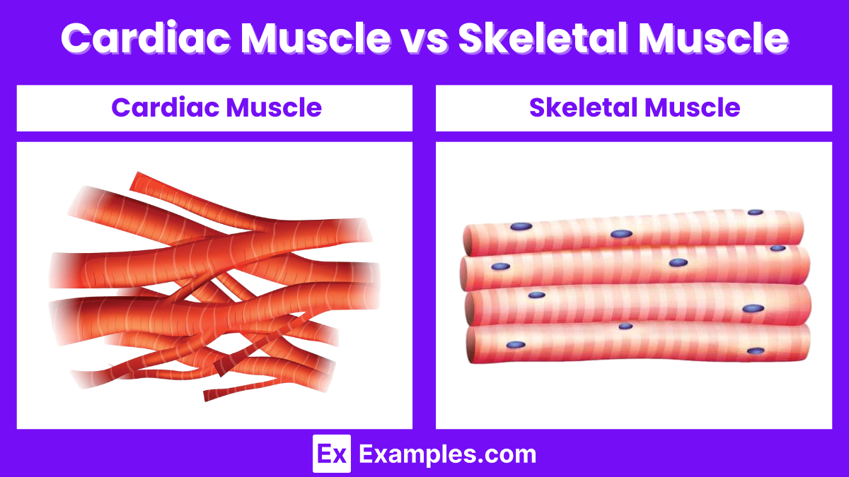
Cardiac muscle and skeletal muscle play crucial roles in the body. Cardiac muscle, found only in the heart, enables the heart to pump blood throughout the body. Skeletal muscle, attached to bones, allows for voluntary movements and supports posture. Both muscle types have unique structures and functions that distinguish them. Understanding these differences helps in comprehending how the muscular system operates and maintains overall health.
Cardiac Muscle
Cardiac muscle, also known as myocardium, is a specialized type of muscle found only in the heart. It is responsible for pumping blood throughout the body, ensuring the delivery of oxygen and nutrients to tissues while removing waste products.
Structure of Cardiac Muscle
Cellular Composition
Cardiac muscle cells, or cardiomyocytes, have unique structural features that distinguish them from skeletal and smooth muscle cells.
- Striated Appearance: Like skeletal muscle, cardiac muscle cells are striated, meaning they have a banded appearance due to the organized arrangement of actin and myosin filaments.
- Intercalated Discs: These are specialized connections between cardiomyocytes. Intercalated discs contain gap junctions and desmosomes, allowing for rapid electrical conduction and strong mechanical attachment between cells.
- Single Nucleus: Each cardiac muscle cell typically contains a single, centrally located nucleus, though some cells may have two nuclei.
- Branching Structure: Cardiomyocytes are branched, forming a complex, interconnected network that facilitates the coordinated contraction of the heart muscle.
Function of Cardiac Muscle
Contraction Mechanism
The contraction of cardiac muscle is similar to that of skeletal muscle, involving the sliding filament theory.
- Action Potential Initiation: The sinoatrial (SA) node, the heart’s natural pacemaker, generates an electrical impulse.
- Calcium Ion Release: This impulse travels through the atria, causing the release of calcium ions from the sarcoplasmic reticulum into the cytoplasm.
- Cross-Bridge Formation: Calcium ions bind to troponin, causing a conformational change in tropomyosin and exposing binding sites on actin filaments.
- Sliding Filaments: Myosin heads bind to actin, forming cross-bridges and pulling the actin filaments toward the center of the sarcomere, resulting in contraction.
Electrical Conduction System
The heart’s electrical conduction system ensures that the cardiac muscle contracts in a coordinated manner.
- Sinoatrial (SA) Node: Located in the right atrium, it initiates the electrical impulse.
- Atrioventricular (AV) Node: The impulse travels to the AV node, which delays the signal before passing it to the ventricles.
- Bundle of His and Purkinje Fibers: The impulse travels through the bundle of His and Purkinje fibers, causing the ventricles to contract.
Role in Health and Disease
Normal Function
In a healthy heart, cardiac muscle efficiently pumps blood, maintaining circulation and supporting overall bodily function.
Pathological Conditions
- Myocardial Infarction (Heart Attack): Blockage of coronary arteries can lead to the death of cardiac muscle cells.
- Cardiomyopathy: Diseases of the heart muscle can weaken its ability to pump blood effectively.
- Arrhythmias: Abnormal electrical activity can lead to irregular heartbeats, affecting the coordination of cardiac muscle contraction.
Skeletal Muscle
Skeletal muscle is a type of muscle tissue that is attached to bones and is responsible for voluntary movements of the body. It is also involved in maintaining posture, stabilizing joints, and generating heat through muscle contractions.
Structure of Skeletal Muscle
Cellular Composition
Skeletal muscle fibers, or myocytes, are long, cylindrical cells that are multinucleated. The following are key structural components of skeletal muscle:
- Sarcolemma: The cell membrane of a muscle fiber.
- Sarcoplasm: The cytoplasm of a muscle fiber, containing organelles, myofibrils, and myoglobin.
- Myofibrils: Rod-like units within muscle fibers composed of repeating sections called sarcomeres, which are the basic functional units of muscle contraction.
- Sarcomeres: Segments of myofibrils containing actin (thin filaments) and myosin (thick filaments) arranged in a specific pattern, giving skeletal muscle its striated appearance.
Connective Tissue Layers
- Endomysium: Surrounds individual muscle fibers.
- Perimysium: Surrounds bundles of muscle fibers called fascicles.
- Epimysium: Encloses the entire muscle, providing structural integrity and support.
Function of Skeletal Muscle
Contraction Mechanism
Skeletal muscle contraction is governed by the sliding filament theory, which involves the following steps:
- Neuromuscular Junction Activation: An action potential from a motor neuron releases acetylcholine into the synaptic cleft, triggering an action potential in the muscle fiber.
- Calcium Ion Release: The action potential travels along the sarcolemma and down T-tubules, causing the sarcoplasmic reticulum to release calcium ions.
- Cross-Bridge Formation: Calcium ions bind to troponin, causing tropomyosin to move away from actin’s binding sites, allowing myosin heads to attach to actin.
- Power Stroke: Myosin heads pivot, pulling actin filaments toward the center of the sarcomere, leading to muscle contraction.
- Cross-Bridge Detachment: ATP binds to myosin, causing it to release actin and re-cock for another cycle.
Types of Skeletal Muscle Fibers
Type I (Slow-Twitch) Fibers
- Characteristics: High endurance, slow contraction speed, high mitochondrial density.
- Function: Suited for endurance activities such as long-distance running.
Type II (Fast-Twitch) Fibers
- Characteristics: Low endurance, fast contraction speed, lower mitochondrial density.
- Subtypes: Type IIa (intermediate), Type IIb (rapid, powerful contractions).
- Function: Suited for short bursts of power and strength activities such as sprinting and weightlifting.
Role in Health and Disease
Normal Function
- Movement: Enables movement of the body and its parts by contracting and relaxing in response to neural signals.
- Posture and Stability: Maintains posture and stabilizes joints.
- Heat Production: Generates heat through muscle contractions, helping to maintain body temperature.
Pathological Conditions
- Muscular Dystrophy: A group of genetic disorders causing progressive muscle weakness and degeneration.
- Myasthenia Gravis: An autoimmune disorder that impairs communication between nerves and muscles, leading to muscle weakness.
- Rhabdomyolysis: A condition involving the breakdown of muscle tissue, releasing damaging proteins into the bloodstream.
Differences between Cardiac Muscle and Skeletal Muscle
| Feature | Cardiac Muscle | Skeletal Muscle |
|---|---|---|
| Location | Found only in the heart | Attached to bones throughout the body |
| Control | Involuntary | Voluntary |
| Cell Shape | Branched and cylindrical | Long and cylindrical |
| Nucleus | Usually one central nucleus | Multiple nuclei per cell, located peripherally |
| Striations | Present | Present |
| Intercalated Discs | Present, allowing synchronized contraction | Absent |
| Contraction Speed | Moderate | Fast |
| Fatigue Resistance | High, rarely fatigues | Varies, can fatigue depending on activity |
| Energy Supply | Primarily aerobic respiration | Both aerobic and anaerobic respiration |
| Function | Pumps blood throughout the body | Enables body movement and posture |
| Regeneration Ability | Limited | Moderate |
| T-tubules | Wider and fewer | Narrow and numerous |
| Sarcoplasmic Reticulum | Less extensive | Highly developed |
| Innervation | Autonomic nervous system | Somatic nervous system |
| Contraction Mechanism | Coordinated contraction through pacemaker cells | Contraction initiated by motor neurons |
| Presence of Myofibrils | Present | Present |
| Calcium Source for Contraction | Extracellular fluid and sarcoplasmic reticulum | Mainly from sarcoplasmic reticulum |
| Role in Homeostasis | Maintains blood circulation, thus crucial for homeostasis | Contributes to movement and heat production |
Similarities Between Cardiac Muscle and Skeletal Muscle
Cardiac muscle and skeletal muscle are two types of muscle tissues in the human body that share several similarities due to their structural and functional characteristics. Understanding these similarities can provide insights into their roles in movement and bodily functions. Below are the key similarities between cardiac muscle and skeletal muscle:
Structural Similarities
- Striations
- Both cardiac and skeletal muscles exhibit a striated appearance under a microscope. This striation is due to the regular arrangement of actin and myosin filaments in sarcomeres, the basic functional units of muscle fibers.
- Sarcomeres
- Both types of muscles have sarcomeres, which are the repeating units that create the striated pattern. Sarcomeres are responsible for muscle contraction and are composed of thin (actin) and thick (myosin) filaments.
- T-tubules
- Cardiac and skeletal muscles both contain T-tubules (transverse tubules). These invaginations of the cell membrane help in the rapid transmission of action potentials, ensuring coordinated contractions.
- Sarcoplasmic Reticulum
- Both muscle types possess a sarcoplasmic reticulum, an organelle that stores and releases calcium ions (Ca²⁺). Calcium release from the sarcoplasmic reticulum is essential for muscle contraction in both cardiac and skeletal muscles.
Functional Similarities
- Excitation-Contraction Coupling
- Both cardiac and skeletal muscles rely on excitation-contraction coupling, a process where an electrical signal (action potential) leads to muscle contraction. This involves the release of calcium ions and the interaction of actin and myosin filaments.
- ATP Dependence
- The contraction of both muscle types is ATP-dependent. ATP provides the necessary energy for the interaction between actin and myosin filaments, leading to muscle contraction.
- Use of Myoglobin
- Both cardiac and skeletal muscles contain myoglobin, a protein that binds oxygen. Myoglobin helps in the storage and transport of oxygen within muscle cells, crucial for sustained muscle activity and endurance.
What are the main differences between cardiac and skeletal muscle?
Cardiac muscle is involuntary and striated, found in the heart. Skeletal muscle is voluntary and striated, attached to bones for movement.
Where is cardiac muscle located?
Cardiac muscle is located exclusively in the heart, enabling it to pump blood throughout the body.
Where is skeletal muscle found?
Skeletal muscle is attached to bones via tendons, facilitating body movement and posture.
How does cardiac muscle function?
Cardiac muscle contracts involuntarily, rhythmically pumping blood through the heart’s chambers and into the circulatory system.
How does skeletal muscle function?
Skeletal muscle contracts voluntarily, controlled by the nervous system, enabling body movements and maintaining posture.
What is the structure of cardiac muscle?
Cardiac muscle cells are branched, interconnected, and contain one or two nuclei, with intercalated discs for synchronized contraction.
What is the structure of skeletal muscle?
Skeletal muscle cells are long, cylindrical, and multinucleated, organized into fibers with a striated appearance.
Do cardiac and skeletal muscles regenerate?
Cardiac muscle has limited regenerative capacity, while skeletal muscle can regenerate to some extent through satellite cells.
What are intercalated discs in cardiac muscle?
Intercalated discs are specialized connections between cardiac muscle cells that facilitate coordinated contractions.
How is skeletal muscle controlled?
Skeletal muscle is controlled by the somatic nervous system, allowing voluntary movement.
Share :
