Centrioles are primarily involved in:
Photosynthesis
Cellular respiration
Cell division
Protein synthesis


Dive into the fascinating world of centrioles with our comprehensive guide! These cylindrical structures play a pivotal role in cell division, particularly in the formation of spindle fibers that separate chromosomes. Through detailed examples, we’ll explore their structure, function, and significance in both animal cells and the wider biological landscape. This guide is tailored for students, educators, and biology enthusiasts seeking to deepen their understanding of cellular biology. With a focus on clarity and accessibility, we make complex concepts approachable, enriching your knowledge of cell biology’s intricate mechanisms.
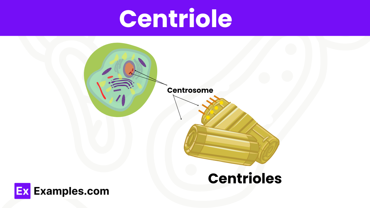
Centrioles are cylindrical structures that are found in most eukaryotic cells, though they are absent in higher plants and most fungi. Typically, a centriole is composed of a short cylinder of microtubules arranged in a circle with a pattern of nine groups of microtubules. Each group contains three microtubules, and this structure is referred to as a “triplet.” The two centrioles in a cell are positioned perpendicular to each other within a region known as the centrosome, which acts as a major microtubule-organizing center and plays a critical role in the spatial arrangement of the cell’s cytoskeleton.
Centrioles are cylindrical structures found in most eukaryotic cells, except for most fungi and all plants. They are key components of the cytoskeleton and play a crucial role in the process of cell division. A centriole is typically composed of nine sets of microtubule triplets arranged in a cylinder. Each microtubule is a hollow tube made up of tubulin proteins. The arrangement of these triplets gives the centriole a distinct, nine-fold symmetry. The centrioles are usually found in pairs and positioned perpendicular to each other within a region of the cell known as the centrosome. This organization is crucial for their role in cell division, particularly during the formation of the mitotic spindle, which is essential for distributing chromosomes into daughter cells.

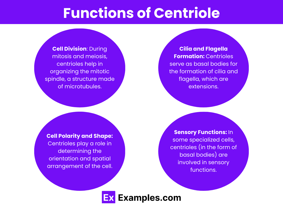
Centrioles play several vital roles in the cell:
Centrioles are absent in most plant cells. Plants have a different mechanism for cell division and do not form centrioles or a classical centrosome. Instead of centrioles, plant cells organize their microtubules using other structures and proteins to form the pre-prophase band, spindle, and phragmoplast during cell division. This difference highlights the evolutionary divergence in the mechanisms of cell division between plant and animal cells. However, some lower plants and algae do have centriole-like structures that play roles similar to those in animal cells, particularly in the formation of flagella.
Centriole duplication is a highly regulated process that occurs once per cell cycle to ensure that each daughter cell inherits a complete centrosome. This process begins in the S phase (DNA synthesis phase) of the cell cycle and is completed by the end of G2 phase, just before mitosis begins. Duplication initiates with the formation of a procentriole near the base of each pre-existing centriole. The procentrioles then elongate, maturing into new centrioles that remain perpendicular to their parental centrioles. The duplication cycle is tightly coordinated with the cell cycle to prevent the formation of extra centrioles.
Centrioles are crucial for cell division, helping organize the mitotic spindle and ensuring accurate chromosome segregation.
A centriole resembles a small, cylindrical structure, composed of nine triplets of microtubules arranged in a ring.
Centrioles were first discovered by Edouard Van Beneden in 1883, during his research on the cell division process.
The Golgi apparatus is a crucial cellular organelle responsible for modifying, sorting, and packaging proteins and lipids for secretion or use within the cell. Acting as the cell’s post office, it ensures that cellular products are correctly modified and sent to their proper destinations, supporting cell maintenance, growth, and communication.
Text prompt
Add Tone
10 Examples of Public speaking
20 Examples of Gas lighting
Centrioles are primarily involved in:
Photosynthesis
Cellular respiration
Cell division
Protein synthesis
Centrioles are found in which type of cells?
Plant cells only
Animal cells only
Both plant and animal cells
Bacterial cells only
What is the structure of a centriole?
A single circular DNA
A double helix
A cylindrical structure made of microtubules
A flat membrane
What is the primary function of centrioles in animal cells?
Photosynthesis
Protein synthesis
Cell division
Detoxification
What is the primary function of centrioles in animal cells?
Photosynthesis
Protein synthesis
Cell division
Energy production
Centrioles are composed primarily of:
DNA
Lipids
Microtubules
RNA
Where are centrioles typically located in animal cells?
Nucleus
Cytoplasm
Near the Golgi apparatus
In the centrosome
How many centrioles are found in a typical centrosome?
One
Two
Three
Four
Which phase of the cell cycle do centrioles duplicate?
G1 phase
S phase
G2 phase
M phase
Centrioles are absent in which of the following cell types?
Animal cells
Plant cells
Fungal cells
Bacterial cells
Before you leave, take our quick quiz to enhance your learning!

