Chromosome – Definition, Structure, Functions, Types, In Humans

Chromosome – Definition, Structure, Functions, Types, In Humans
Chromosomes hold the blueprint for all living organisms. These thread-like structures reside in the nucleus of each cell and carry genetic information in the form of DNA. They play a critical role in heredity, influencing traits passed from parents to offspring. Understanding chromosomes is essential for comprehending the fundamentals of genetics, evolution, and cellular function. Each species has a specific number of chromosomes, which determine the complexity and diversity of life forms on Earth. This article delves into the structure, function, and significance of chromosomes in biological systems.
What is a Chromosome?
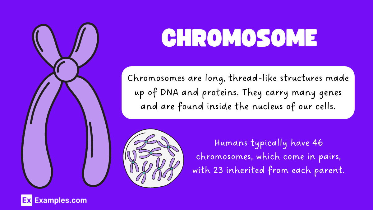
A chromosome is a long, thread-like structure made of DNA and proteins. It is found in the nucleus of eukaryotic cells and contains the genetic information necessary for the development, functioning, and reproduction of an organism. Chromosomes ensure the accurate distribution of genetic material during cell division.
Examples of Chromosomes
Human Chromosomes
Humans have 23 pairs of chromosomes, totaling 46. These include 22 pairs of autosomes and one pair of sex chromosomes (XX for females and XY for males). Each chromosome carries numerous genes that determine various traits and functions.
Fruit Fly Chromosomes
Fruit flies (Drosophila melanogaster) possess 8 chromosomes. Their genetic simplicity makes them ideal for genetic research and studies on inheritance patterns.
Plant Chromosomes
Arabidopsis thaliana, a model organism in plant biology, has 5 pairs of chromosomes. Its relatively small genome allows for extensive genetic research and manipulation.
Bacterial Chromosomes
Escherichia coli, a common bacterium, typically contains a single circular chromosome. This chromosome carries all the genetic information needed for the bacterium’s survival and reproduction.
Mouse Chromosomes
Mice have 20 pairs of chromosomes. These chromosomes are crucial for studying mammalian genetics, as mice share a significant amount of genetic similarity with humans.
Yeast Chromosomes
Saccharomyces cerevisiae, a type of yeast, has 16 chromosomes. Its simple eukaryotic structure provides insights into fundamental cellular processes.
Functions of Chromosomes
Gene Regulation
Chromosomes play a vital role in controlling the expression of genes. They organize DNA in a way that allows genes to be turned on or off as needed, ensuring that the right proteins are produced at the right times.
Inheritance
Chromosomes are responsible for transmitting genetic information from parents to offspring. During reproduction, chromosomes from each parent combine to form a complete set, providing the genetic blueprint for the new organism.
Cell Division
Chromosomes ensure the accurate distribution of genetic material during cell division. This process occurs in two main types:
- Mitosis: Produces two identical daughter cells, each with the same number of chromosomes as the parent cell. This is crucial for growth, development, and tissue repair.
- Meiosis: Produces four genetically diverse daughter cells, each with half the number of chromosomes as the parent cell. This is essential for sexual reproduction and genetic diversity.
Maintenance of Genetic Stability
Chromosomes help maintain the integrity and stability of an organism’s genome. They protect DNA from damage and ensure that genetic information is accurately copied and passed on during cell division.
Structure of Chromosome
1. Chromatin
Chromosomes are made up of chromatin, a complex of DNA and proteins. Chromatin exists in two forms:
- Euchromatin: This is less condensed and contains genes that are actively being transcribed.
- Heterochromatin: This is more condensed and typically contains genes that are not actively transcribed.
2. DNA and Histones
DNA Structure:
- Double Helix: The basic structure of DNA is a double helix, where two strands of nucleotides are twisted around each other.
- Nucleotides: Each nucleotide is composed of a sugar, a phosphate group, and a nitrogenous base (adenine, thymine, cytosine, or guanine).
Histones:
- Nucleosomes: DNA wraps around histone proteins to form nucleosomes, the basic unit of chromatin. Each nucleosome consists of DNA wrapped around a core of eight histone proteins.
- Linker DNA: Nucleosomes are connected by linker DNA, which is bound by another histone protein, H1, to help further compact the DNA.
3. Chromosome Structure
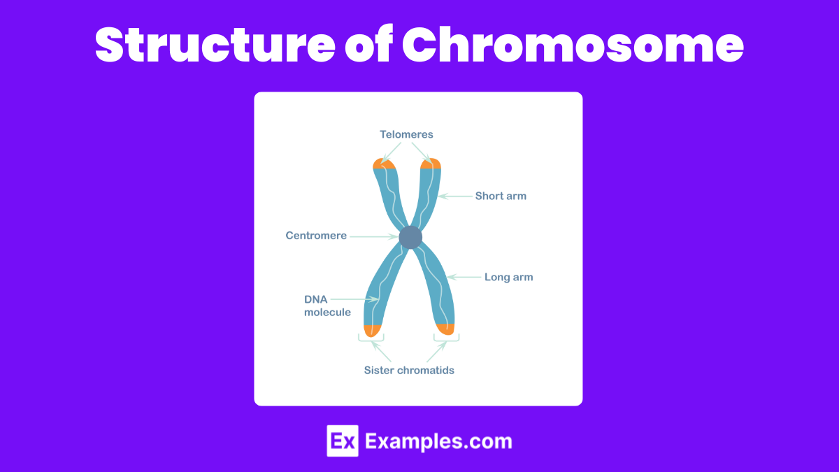
Centromere:
- Location: The centromere is a constricted region of the chromosome that divides it into two arms.
- Function: It is essential for the movement of chromosomes during cell division as it is the attachment site for spindle fibers.
Telomeres:
- Location: Telomeres are repetitive nucleotide sequences at the ends of chromosomes.
- Function: They protect the chromosome ends from deterioration and prevent fusion with other chromosomes.
Arms:
- p Arm: The shorter arm of the chromosome.
- q Arm: The longer arm of the chromosome.
4. Chromatid
Definition:
- Sister Chromatids: After DNA replication, each chromosome consists of two identical sister chromatids, joined at the centromere.
Function:
- Cell Division: During mitosis and meiosis, sister chromatids are separated and distributed to daughter cells, ensuring each new cell receives an identical set of chromosomes.
5. Chromosome Classification
Chromosomes can be classified based on the position of the centromere:
- Metacentric: The centromere is in the middle, resulting in two arms of equal length.
- Submetacentric: The centromere is slightly off-center, leading to one arm being longer than the other.
- Acrocentric: The centromere is close to one end, creating one very long arm and one very short arm.
- Telocentric: The centromere is at the very end of the chromosome (not found in humans).
6. Karyotype
Definition:
- Karyotype: A karyotype is a complete set of chromosomes in a species or an individual organism, often displayed in a standardized format showing the number, size, and shape of each chromosome type.
Use:
- Genetic Analysis: Karyotyping is used to diagnose genetic disorders and to study chromosomal abnormalities.
What are centromeres?
Definition and Structure: Centromeres are specialized regions of a chromosome that play a pivotal role during cell division. They are the points where sister chromatids (the identical copies of a chromosome) are most closely attached to each other after DNA replication.
Functions:
- Kinetochore Formation: The centromere is the site where the kinetochore, a complex of proteins, forms. The kinetochore is essential for the attachment of spindle fibers during mitosis and meiosis.
- Chromosome Segregation: During cell division, spindle fibers attach to the kinetochores and pull the sister chromatids apart, ensuring that each daughter cell receives an identical set of chromosomes.
- Genetic Stability: By ensuring accurate chromosome segregation, centromeres help maintain genetic stability, preventing aneuploidy (abnormal number of chromosomes), which can lead to diseases like cancer.
Types of Centromeres:
- Point Centromeres: Found in some species like yeast, these are small and specific DNA sequences.
- Regional Centromeres: Found in most eukaryotes, these are larger and less specific, containing repetitive DNA sequences.
What are telomeres?
Definition and Structure: Telomeres are repetitive nucleotide sequences at the ends of chromosomes. In humans, the sequence is typically “TTAGGG” repeated thousands of times. They are protected by a complex of proteins known as shelterin.
Functions:
- Protection of Chromosome Ends: Telomeres prevent the ends of chromosomes from being recognized as broken DNA, which would otherwise trigger DNA repair mechanisms leading to chromosome end-to-end fusions.
- Prevention of DNA Loss: Every time a cell divides, a small portion of the DNA at the end of the chromosome is lost. Telomeres ensure that this loss does not affect vital genes.
- Regulation of Cell Aging: Telomeres shorten with each cell division. When they become too short, the cell can no longer divide and becomes senescent or undergoes apoptosis (programmed cell death). This mechanism limits the number of times a cell can divide, contributing to the aging process.
Telomerase and Telomere Maintenance:
- Telomerase: An enzyme called telomerase can extend the length of telomeres by adding nucleotide sequences. Telomerase activity is high in germ cells, stem cells, and cancer cells, but low in most somatic cells.
- Telomere Length and Disease: Abnormally short telomeres are associated with aging and age-related diseases, while high telomerase activity in somatic cells can lead to cancer by allowing cells to divide indefinitely.
Types of Chromosomes
Chromosomes can be classified in several ways based on their structure, function, and number in organisms. Below is an overview of the different types of chromosomes:
Based on Centromere Position
Chromosomes can be classified according to the position of their centromere, which affects the relative lengths of their arms:
a. Metacentric Chromosomes
- Definition: The centromere is located near the middle, producing two arms of approximately equal length.
- Example: Human chromosome 1.
b. Submetacentric Chromosomes
- Definition: The centromere is slightly off-center, resulting in one arm being slightly longer than the other.
- Example: Human chromosome 4.
c. Acrocentric Chromosomes
- Definition: The centromere is significantly off-center, creating one very long arm and one very short arm.
- Example: Human chromosome 13.
d. Telocentric Chromosomes
- Definition: The centromere is located at the very end of the chromosome, forming only one arm.
- Example: Not found in humans, but present in some species such as mice.
Based on Chromosome Function
a. Autosomes
- Definition: Chromosomes that are not directly involved in determining the sex of an individual.
- Number in Humans: Humans have 22 pairs of autosomes.
- Function: Carry the majority of an organism’s genetic information and are involved in a wide range of bodily functions.
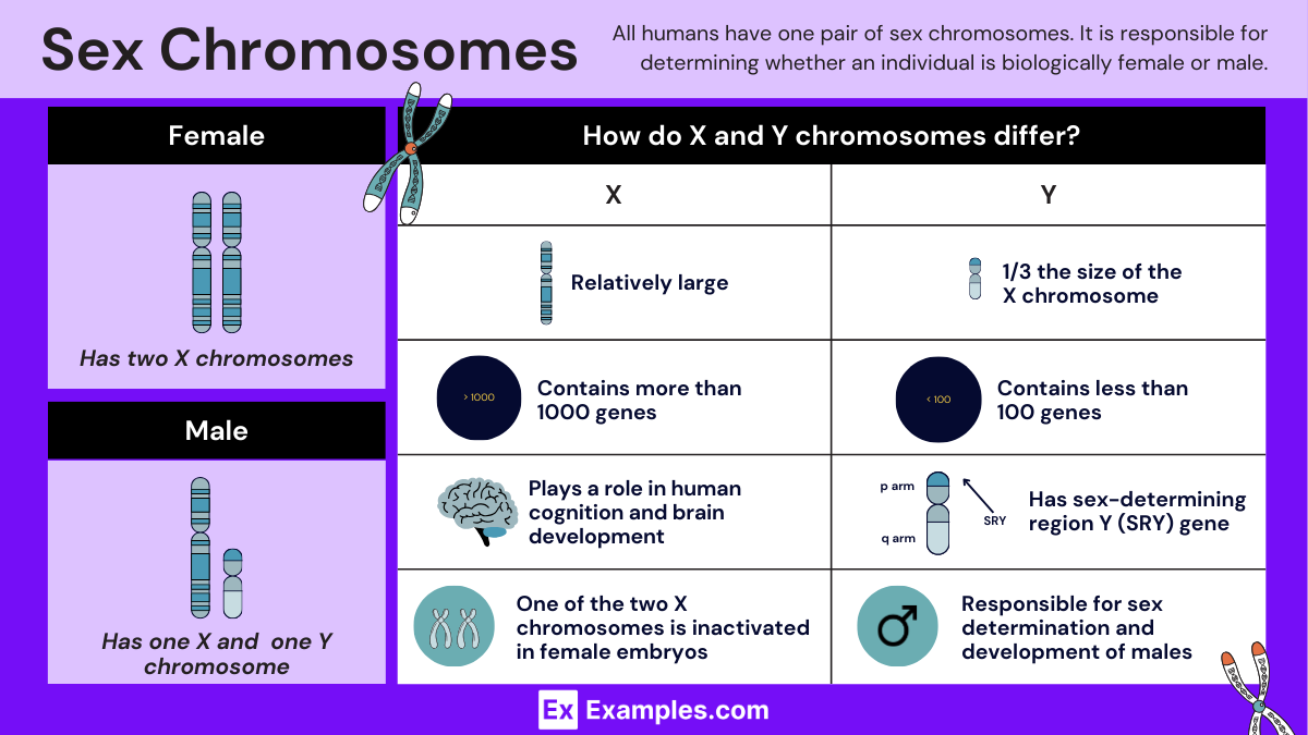
b. Sex Chromosomes
- Definition: Chromosomes that determine the sex of an individual.
- Types in Humans: X and Y chromosomes.
- Number in Humans: Humans have one pair of sex chromosomes (XX for females, XY for males).
Based on Chromosome Number
a. Haploid (n)
- Definition: Cells that contain a single set of chromosomes.
- Example: Gametes (sperm and egg cells) in humans have 23 chromosomes each.
b. Diploid (2n)
- Definition: Cells that contain two sets of chromosomes, one from each parent.
- Example: Somatic (body) cells in humans have 46 chromosomes (23 pairs).
c. Polyploid
- Definition: Cells that contain more than two sets of chromosomes.
- Example: Many plant species, such as wheat (hexaploid, 6n), are polyploid.
Special Types of Chromosomes
a. B Chromosomes (Accessory Chromosomes)
- Definition: Extra chromosomes found in some species, not essential for normal growth and development.
- Function: Their function is often unclear, but they may play a role in genetic diversity and evolution.
b. Giant Chromosomes (Polytene Chromosomes)
- Definition: Chromosomes that have undergone multiple rounds of DNA replication without cell division, resulting in large, visible chromosomes.
- Example: Found in the salivary glands of Drosophila (fruit fly) larvae.
c. Lampbrush Chromosomes
- Definition: Extended, looped chromosomes that are active in transcription and visible during the oocyte development of some vertebrates, particularly amphibians.
- Function: Facilitates high levels of gene transcription needed during oocyte development.
Do all living things have the same types of chromosomes?
Not all living things have the same types of chromosomes. The structure, number, and organization of chromosomes vary widely among different species. For example, humans have 23 pairs of linear chromosomes, while bacteria typically have a single circular chromosome. Plants and animals can have varying numbers of chromosomes, often differing in size and shape. Additionally, some organisms, like certain plants and fungi, have multiple sets of chromosomes (polyploidy), whereas others, like viruses, possess minimal genetic material in simple forms. This diversity reflects the evolutionary adaptations and complexities of each organism’s genetic makeup.
Chromosomes in Humans
Humans have a total of 46 chromosomes, which are organized into 23 pairs. These chromosomes are found in the nucleus of every cell and play a crucial role in storing and transmitting genetic information.
Breakdown of Chromosome Pairs
- Autosomes
- 22 pairs of chromosomes are autosomes. These are numbered from 1 to 22 based on their size, with chromosome 1 being the largest and chromosome 22 the smallest.
- Autosomes are identical in both males and females and carry the bulk of an individual’s genetic information.
- Sex Chromosomes
- 1 pair of chromosomes are sex chromosomes, which determine an individual’s sex.
- Females have two X chromosomes (XX).
- Males have one X chromosome and one Y chromosome (XY).
Do males have different chromosomes than females?
Yes, males and females have different sets of sex chromosomes, which determine their biological sex. While both sexes share the same autosomes, the sex chromosomes vary.
- Autosomes
- Both males and females have 22 pairs of autosomes, which are identical in both sexes and carry most of the genetic information.
- Sex Chromosomes
- Females have two X chromosomes (XX).
- Males have one X chromosome and one Y chromosome (XY).
Differences in Genetic Contribution
- Females (XX)
- Both X chromosomes contribute equally to genetic traits.
- During meiosis (the process of forming eggs), females pass on one of their X chromosomes to their offspring.
- Males (XY)
- The X chromosome comes from the mother, and the Y chromosome comes from the father.
- During meiosis (the process of forming sperm), males can pass on either their X or Y chromosome, determining the sex of the offspring:
- X chromosome from the male results in a female offspring (XX).
- Y chromosome from the male results in a male offspring (XY).
| Feature | Females (XX) | Males (XY) |
|---|---|---|
| Total Chromosomes | 46 (23 pairs) | 46 (23 pairs) |
| Autosomes | 22 pairs (numbered 1-22) | 22 pairs (numbered 1-22) |
| Sex Chromosomes | Two X chromosomes (XX) | One X and one Y chromosome (XY) |
| X Chromosome | Present in two copies | Present in one copy |
| Y Chromosome | Absent | Present in one copy |
| Sex Determination | Does not determine sex of offspring | Determines sex of offspring (X or Y) |
Duplication of Chromosomes
Chromosome duplication refers to the process by which a cell makes an exact copy of each chromosome, resulting in two identical sets. This occurs during the S phase (Synthesis phase) of the cell cycle, before a cell divides.
Phases of Chromosome Duplication
- Interphase:
- G1 Phase (Gap 1): The cell grows and synthesizes proteins necessary for DNA replication.
- S Phase (Synthesis): Each chromosome is duplicated. The DNA unzips and new nucleotides are added to form two identical sister chromatids.
- G2 Phase (Gap 2): The cell continues to grow and prepares for mitosis. The duplicated chromosomes are checked for errors.
- Mitosis:
- Prophase: Chromosomes condense and become visible. The nuclear envelope breaks down.
- Metaphase: Chromosomes align at the cell’s equator.
- Anaphase: Sister chromatids are pulled apart to opposite poles of the cell.
- Telophase: Nuclear envelopes reform around the two sets of chromosomes.
- Cytokinesis: The cell divides into two daughter cells, each with a complete set of chromosomes.
Common Errors in Chromosome Duplication
- Mutations: Changes in the DNA sequence can occur during duplication.
- Aneuploidy: Abnormal number of chromosomes, leading to conditions like Down syndrome.
- Replication Stress: Faulty replication machinery can cause DNA damage and genomic instability.
Examples of Chromosome Duplication
- Cancer Cells: Often exhibit abnormal chromosome duplication, leading to genomic instability.
- Polyploidy in Plants: Many plants, such as wheat, have multiple sets of chromosomes due to duplication events, contributing to their diversity and adaptability.
| Phase | Key Events |
|---|---|
| G1 Phase | Cell growth, preparation for DNA replication |
| S Phase | DNA synthesis, chromosome duplication |
| G2 Phase | Cell growth, preparation for mitosis, error checking |
| Prophase | Chromosomes condense, nuclear envelope breaks down |
| Metaphase | Chromosomes align at the equator |
| Anaphase | Sister chromatids separate to opposite poles |
| Telophase | Nuclear envelopes reform |
| Cytokinesis | Cell divides into two daughter cells |
Discovery of Chromosomes
Early Observations
- Anton van Leeuwenhoek (1670s):
- Using one of the earliest microscopes, Leeuwenhoek observed sperm cells and noted their structure, laying the groundwork for cellular biology.
- Robert Hooke (1665):
- Coined the term “cell” after observing cork cells under a microscope, which contributed to the study of cell structure.
Advancements in Microscopy
- Early 19th Century:
- Improvements in microscope technology allowed scientists to observe cells in greater detail, leading to discoveries about cell division and internal structures.
Key Discoveries
- Friedrich Miescher (1869):
- Discovered a substance he called “nuclein” (now known as DNA) in the nuclei of white blood cells. This substance was later understood to be associated with chromosomes.
- Walther Flemming (1879):
- Observed and described the process of mitosis in animal cells. Flemming used aniline dyes to stain cells and observed thread-like structures, which he named “chromatin.”
- Edouard Van Beneden (1883):
- Discovered that the number of chromosomes is constant in the cells of a species. He also observed meiosis, noting the reduction in chromosome number in gametes.
The Term “Chromosome”
- Heinrich Wilhelm Gottfried von Waldeyer-Hartz (1888):
- Coined the term “chromosome” (from the Greek words “chroma,” meaning color, and “soma,” meaning body) to describe the stained, thread-like structures observed during cell division.
Chromosome Theory of Inheritance
- Walter Sutton and Theodor Boveri (1902):
- Independently proposed the chromosome theory of inheritance, suggesting that chromosomes carry the genetic material and are responsible for the transmission of traits from one generation to the next.
- Thomas Hunt Morgan (1910s):
- Conducted experiments with fruit flies (Drosophila melanogaster) and provided definitive evidence for the chromosome theory of inheritance. He demonstrated that genes are located on chromosomes and that specific traits are linked to specific chromosomes.
How are chromosomes inherited?
Chromosomes are inherited through a process called meiosis, which occurs in the reproductive cells of organisms. During meiosis, the chromosome number is halved, resulting in gametes (sperm and egg cells) that contain only one set of chromosomes, or 23 in humans. When fertilization occurs, a sperm cell and an egg cell combine to form a zygote with a full set of 46 chromosomes—23 from the mother and 23 from the father. This ensures that offspring inherit a mix of genetic material from both parents, contributing to genetic diversity.
Abnormalities of the chromosome
Chromosomal abnormalities occur when there is an irregularity in the number or structure of chromosomes. These abnormalities can lead to various genetic disorders and conditions. The main types of chromosomal abnormalities include:
1. Numerical Abnormalities:
- Trisomy: This occurs when there is an extra chromosome in the cells. An example is Down syndrome, caused by an extra copy of chromosome 21.
- Monosomy: This is when a chromosome is missing. Turner syndrome, where females have only one X chromosome, is a classic example.
2. Structural Abnormalities:
- Deletions: A portion of the chromosome is missing or deleted. An example is Cri-du-chat syndrome, which involves a deletion on chromosome 5.
- Duplications: A portion of the chromosome is duplicated, resulting in extra genetic material. Charcot-Marie-Tooth disease type 1A is caused by a duplication of a segment on chromosome 17.
- Translocations: A segment of one chromosome is transferred to another chromosome. This can be balanced (no genetic material is lost or gained) or unbalanced (some genetic material is extra or missing). Chronic myelogenous leukemia is associated with a translocation between chromosomes 9 and 22.
- Inversions: A portion of the chromosome breaks off, flips around, and reattaches, reversing the gene sequence. Although inversions often do not cause problems, they can lead to reproductive issues.
- Rings: A chromosome forms a ring structure due to deletions in the telomeres, which are the ends of chromosomes. This can lead to various developmental and health issues.
Chromosome Conditions and Disorders
Chromosome Number Abnormalities
Chromosome number abnormalities occur when there are extra or missing chromosomes. These abnormalities can be detected using karyotyping.
Down Syndrome (Trisomy 21)
- Description: Down Syndrome is caused by an extra copy of chromosome 21.
- Symptoms: Intellectual disability, characteristic facial features, heart defects, and developmental delays.
- Incidence: About 1 in 700 births.
Turner Syndrome (Monosomy X)
- Description: Turner Syndrome affects females who have only one X chromosome.
- Symptoms: Short stature, infertility, heart defects, and certain learning disabilities.
- Incidence: About 1 in 2,500 female births.
Klinefelter Syndrome (XXY Syndrome)
- Description: Klinefelter Syndrome affects males who have an extra X chromosome (XXY).
- Symptoms: Reduced testosterone levels, infertility, breast development, and learning disabilities.
- Incidence: About 1 in 500 to 1,000 male births.
Chromosome Structure Abnormalities
Chromosome structure abnormalities result from deletions, duplications, inversions, or translocations of chromosome segments.
Cri-du-Chat Syndrome (5p- Syndrome)
- Description: Cri-du-Chat Syndrome is caused by a deletion of a portion of chromosome 5.
- Symptoms: High-pitched cat-like cry, intellectual disability, delayed development, and distinctive facial features.
- Incidence: About 1 in 20,000 to 50,000 births.
Williams Syndrome
- Description: Williams Syndrome is caused by a deletion of approximately 26 genes from chromosome 7.
- Symptoms: Distinctive facial features, cardiovascular problems, intellectual disability, and an outgoing personality.
- Incidence: About 1 in 10,000 births.
Wolf-Hirschhorn Syndrome (4p- Syndrome)
- Description: Wolf-Hirschhorn Syndrome results from a deletion of the distal portion of chromosome 4.
- Symptoms: Intellectual disability, distinct facial features, growth delays, and seizures.
- Incidence: About 1 in 50,000 births.
Sex Chromosome Disorders
Sex chromosome disorders involve abnormalities in the sex chromosomes (X and Y).
Triple X Syndrome (47,XXX)
- Description: Triple X Syndrome affects females who have an extra X chromosome.
- Symptoms: Often mild or no symptoms; may include tall stature, learning disabilities, and delayed speech and language skills.
- Incidence: About 1 in 1,000 female births.
XYY Syndrome (47,XYY)
- Description: XYY Syndrome affects males who have an extra Y chromosome.
- Symptoms: Often mild; may include tall stature, acne, learning difficulties, and behavioral issues.
- Incidence: About 1 in 1,000 male births.
Diagnosis and Treatment
Diagnosis of chromosome disorders typically involves genetic testing such as karyotyping, FISH (fluorescence in situ hybridization), and array CGH (comparative genomic hybridization).
- Prenatal Testing: Amniocentesis and chorionic villus sampling (CVS) can detect chromosome abnormalities before birth.
- Postnatal Testing: Blood tests and other tissue samples are used to diagnose chromosome conditions in children and adults.
Management and Support
While there is no cure for chromosome disorders, management focuses on addressing symptoms and providing supportive care.
- Medical Treatment: May include surgeries for congenital defects, medications for associated health issues, and hormone therapies.
- Therapies: Physical therapy, occupational therapy, speech therapy, and educational support can improve quality of life.
- Support Groups: Connecting with support groups can provide emotional support and resources for families affected by chromosome disorders.
What is a chromosome?
A chromosome is a long DNA molecule with part or all of the genetic material of an organism, essential for inheritance and proper cellular function.
How many chromosomes do humans have?
Humans have 46 chromosomes, arranged in 23 pairs, with one set inherited from each parent.
What is the function of chromosomes?
Chromosomes carry genes that determine traits, regulate cell functions, and guide growth and development.
What are autosomes?
Autosomes are the 22 pairs of chromosomes that are not involved in determining sex, carrying most of the genetic information.
What are sex chromosomes?
Sex chromosomes, X and Y, determine an individual’s sex; females have XX, while males have XY.
What is a karyotype?
A karyotype is an image of an individual’s chromosomes, used to diagnose genetic disorders and abnormalities.
What are homologous chromosomes?
Homologous chromosomes are pairs of chromosomes, one from each parent, that have the same genes at the same loci but possibly different alleles.
What is chromosomal mutation?
Chromosomal mutations are changes in the structure or number of chromosomes, often leading to genetic disorders.
How are chromosomes studied?
Chromosomes are studied using techniques like karyotyping, fluorescence in situ hybridization (FISH), and genomic sequencing.
What is a centromere?
A centromere is the region of a chromosome that links sister chromatids and is crucial during cell division for proper chromosome alignment and separation.
Share :
