Which type of neuron is primarily responsible for transmitting information from sensory receptors to the central nervous system?
Motor neurons
Sensory neurons
Interneurons
Glial cells
of 10

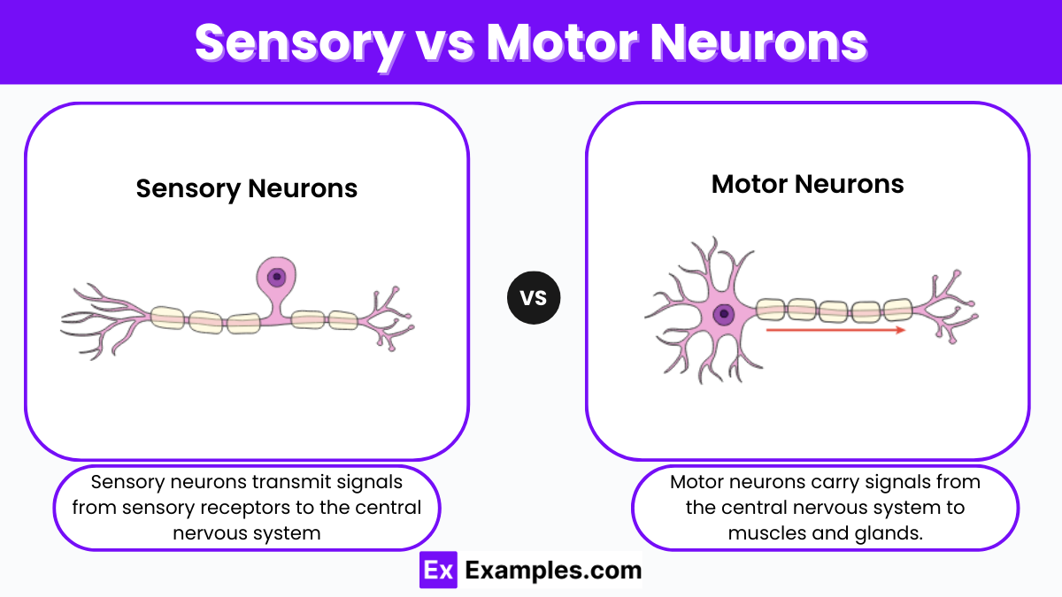
Sensory and motor neurons play crucial roles in the nervous system. Sensory neurons transmit signals from sensory receptors to the central nervous system, allowing us to perceive the world. Motor neurons carry commands from the central nervous system to muscles and glands, enabling movement and responses. Understanding the differences between these two types of neurons helps us grasp how the nervous system processes information and orchestrates actions. This article explores the unique functions, structures, and pathways of sensory and motor neurons, providing a clear comparison to highlight their essential roles in our bodies.
Sensory neurons are specialized nerve cells responsible for converting external stimuli from the organism’s environment into internal electrical impulses. These neurons play a critical role in the sensory system by transmitting information about sensory experiences to the central nervous system (CNS).
Sensory neurons detect and respond to different types of stimuli, such as:
These stimuli are converted into electrical signals through a process called transduction. The sensory neurons then send these signals to the brain or spinal cord, where they are processed and interpreted.
Sensory neurons have a unique structure that allows them to efficiently detect and transmit sensory information. Key components include:
Sensory neurons are crucial for survival and everyday functioning. They help organisms interact with their environment by providing information about potential dangers, aiding in navigation, and facilitating communication.
| Type of Sensory Neuron | Stimuli Detected | Location |
|---|---|---|
| Mechanoreceptors | Touch, pressure, vibration | Skin, joints, muscles |
| Thermoreceptors | Temperature changes | Skin, hypothalamus |
| Nociceptors | Pain | Nearly all body tissues |
| Photoreceptors | Light | Retina of the eyes |
| Chemoreceptors | Chemical stimuli | Taste buds, nasal cavity |
Motor neurons are specialized nerve cells that transmit signals from the central nervous system (CNS) to the muscles and glands in the body, facilitating movement and various physiological responses. These neurons play a critical role in the motor system, enabling voluntary and involuntary actions.
Motor neurons are responsible for conveying instructions from the brain and spinal cord to the effectors (muscles and glands) to initiate movement or secretion. The main functions include:
Motor neurons have a unique structure designed to transmit electrical signals efficiently. Key components include:
Motor neurons are essential for all voluntary and involuntary movements. They enable the body to interact with the environment, perform daily activities, and maintain vital physiological processes.
| Type of Motor Neuron | Function | Location |
|---|---|---|
| Upper Motor Neurons (UMNs) | Transmit signals from the brain to the spinal cord | Motor cortex and brainstem |
| Lower Motor Neurons (LMNs) | Transmit signals from the spinal cord to muscles | Spinal cord and brainstem |
| Somatic Motor Neurons | Control voluntary skeletal muscle movements | Spinal cord, peripheral nerves |
| Autonomic Motor Neurons | Control involuntary muscle movements and gland secretions | Spinal cord, autonomic ganglia |
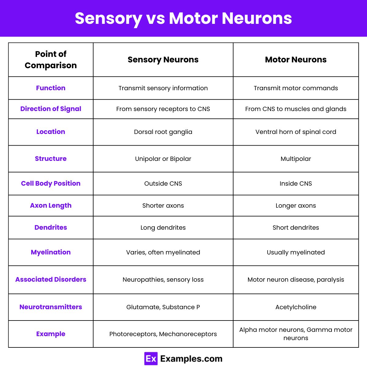
| Feature | Sensory Neurons | Motor Neurons |
|---|---|---|
| Function | Transmit sensory information | Transmit motor commands |
| Direction of Signal | From sensory receptors to CNS | From CNS to effectors (muscles, glands) |
| Location | Found in sensory organs (skin, eyes, ears) | Found in the spinal cord and brain |
| Structure | Typically unipolar or bipolar | Typically multipolar |
| Cell Body Location | In the dorsal root ganglia | In the CNS |
| Dendrites | Few, long dendrites | Numerous, short dendrites |
| Axon Length | Short or long | Usually long |
| Presence of Myelin Sheath | Can be myelinated or unmyelinated | Usually myelinated |
| Neurotransmitters Used | Various (e.g., glutamate) | Mainly acetylcholine |
| Response Type | Afferent (sensory input to CNS) | Efferent (motor output from CNS) |
| Receptor Type | Sensory receptors (mechanoreceptors, photoreceptors) | No specific sensory receptors, synapse on effectors |
| Impulse Origin | Begins at the sensory receptor | Begins in the CNS |
| Synapse Location | Synapse in the CNS | Synapse on muscles or glands |
| Role in Reflex Arc | Carry input from stimulus to spinal cord | Carry output from spinal cord to effector |
| Example | Rods and cones in the retina | Motor neurons in skeletal muscles |
Sensory neurons transmit signals from sensory receptors to the central nervous system, enabling perception of stimuli like touch, pain, temperature, and sound.
Motor neurons carry signals from the central nervous system to muscles or glands, facilitating movement and response actions.
Sensory neurons are located in sensory organs like the skin, eyes, ears, and throughout the body where sensory receptors exist.
Motor neurons are located in the central nervous system, particularly in the spinal cord and brainstem, extending to muscles and glands.
The primary function of sensory neurons is to detect environmental changes and relay this information to the central nervous system.
The primary function of motor neurons is to convey signals from the central nervous system to effectors, causing muscle contraction or gland secretion.
Sensory neurons have limited regeneration capabilities, often relying on peripheral support cells for repair.
Motor neurons have a limited capacity for regeneration, primarily relying on supportive cells and therapies for potential repair.
An example of sensory neuron function is detecting heat from a hot surface and sending pain signals to the brain.
An example of motor neuron function is transmitting signals to leg muscles, enabling walking or running.

Which type of neuron is primarily responsible for transmitting information from sensory receptors to the central nervous system?
Motor neurons
Sensory neurons
Interneurons
Glial cells
What function do motor neurons serve in the nervous system?
They carry information from the brain to muscles and glands.
They transmit sensory information to the brain.
They connect different parts of the brain.
They support and nourish neurons.
Which of the following correctly describes the direction of impulse transmission in sensory neurons?
From muscles to the brain
From the brain to sensory organs
From sensory receptors to the central nervous system
From the central nervous system to muscles
In a reflex action, which type of neuron directly interacts with motor neurons to produce a response?
Sensory neurons
Interneurons
Glial cells
Efferent neurons
Where are the cell bodies of sensory neurons typically located?
In the spinal cord
In the brainstem
In the dorsal root ganglia
In the motor cortex
Which type of neuron is involved in voluntary movements such as writing or walking?
Sensory neurons
Motor neurons
Interneurons
Autonomic neurons
Which of the following statements is true about the sensory neurons' role in the nervous system?
They carry signals away from the central nervous system.
They are responsible for muscle contractions.
They transmit sensory information to the central nervous system.
They regulate internal organ functions.
Which structure typically distinguishes motor neurons from sensory neurons?
Location of the cell body
Length of the axon
Type of neurotransmitter used
Presence of myelin sheath
How do sensory and motor neurons work together to execute a movement?
Sensory neurons transmit the command to move, and motor neurons receive it.
Motor neurons detect stimuli and sensory neurons generate a response.
Sensory neurons detect stimuli, and motor neurons execute the response.
Both types of neurons transmit commands in the same direction.
What role do motor neurons play in the autonomic nervous system?
They regulate voluntary muscle movements.
They control reflex actions.
They manage involuntary functions such as heart rate and digestion.
They process sensory information from the environment.