Which of the following best describes a tendon?
A type of muscle
A connective tissue that attaches muscles to bones
A type of bone
A fluid-filled sac
of 10
Tendons function as dynamic mechanical bridges, facilitating the transfer of muscle force to bones and joints, thus enabling purposeful movements. These complex structures are tailored to the muscles they serve, reflecting specific morphologies and functions. Beyond merely connecting the beginning or end of a muscle, tendons integrate fully with the muscle tissue through the merging of connective layers—epimysium, perimysium, and endomysium—forming a cohesive unit that anchors to bones. This integration allows for a reciprocal relationship where the muscle influences tendon behavior, and the tendon, in turn, shapes the muscle’s functional output.
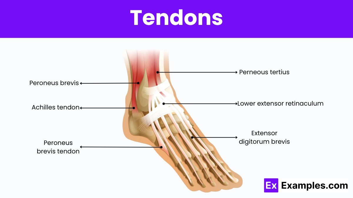
A tendon is a robust, flexible cord of tissue that functions much like a rope, connecting muscles to bones and enabling limb movement. These structures are essential for motion and play a crucial role in safeguarding muscles by absorbing impacts during activities like running and jumping. Found throughout the body, from the head to the toes, tendons are plentiful and diverse in their locations and functions. The largest tendon, the Achilles, links the calf muscle to the heel bone. While tendons are highly resistant to tears, they lack elasticity, making them susceptible to injuries from overstretching and often resulting in prolonged healing times.
Tendons are categorized based on their shape and the specific function they serve in connecting muscles to bones. Understanding these types can help in recognizing their roles in the body’s mechanics and how they contribute to different movements. Here are the primary types of tendons:
1. Long Tendons: These tendons are typically associated with long muscles and are crucial in facilitating movements over large distances. They are slim and cord-like, designed to work with muscles that control fine motor skills. An example of a long tendon is the Achilles tendon, which connects the calf muscles to the heel and is pivotal for running and jumping.
2. Short Tendons: Short tendons are often found in shorter muscles and are usually thicker and wider than long tendons. These tendons are designed for power rather than speed or range of motion. They provide stability and strength in movements. An example can be found in the tendons of the rotator cuff in the shoulder, which help in performing movements like lifting and rotating the arm.
3. Flat Tendons: Flat tendons, or aponeuroses, are broad and flat. They cover a large area and often serve as attachments for sheet-like muscles. These tendons provide support and maintain the position of muscles across a surface. The tendons in the abdominal muscles, which help in flexing and stabilizing the torso, are examples of flat tendons.
4. Sesamoid Tendons: These tendons incorporate small, sesame seed-shaped bones called sesamoids that help in changing the angle of the tendon’s force as it passes to the bone, enhancing the muscle’s mechanical advantage. A well-known sesamoid tendon is the one in the thumb, which helps increase the thumb’s range of motion and gripping power.
The human body contains numerous tendons, each critical for various movements and activities. Here are some of the major tendons that are essential for daily functioning:
1. Achilles Tendon: The Achilles tendon, also known as the calcaneal tendon, is the largest and strongest tendon in the body. It connects the calf muscles (gastrocnemius and soleus) to the heel bone (calcaneus). This tendon is vital for walking, running, and jumping, facilitating the push-off phase that propels the body forward.
2. Patellar Tendon: Located in the knee, the patellar tendon plays a crucial role in the mechanism of the knee joint. It connects the patella (kneecap) to the shin bone (tibia) and works with the muscles of the front thigh (quadriceps) to extend the knee, allowing for movements such as kicking, running, and jumping.
3. Rotator Cuff Tendons: The rotator cuff is a group of four tendons that stabilize the shoulder and allow for a wide range of arm movements. These tendons are:
4. Biceps Tendon: The biceps tendon attaches the biceps muscle to the bones of the shoulder and elbow. It helps in flexing the elbow and rotating the forearm. This tendon is crucial for movements involving lifting and pulling.
5. Quadriceps Tendon: This tendon connects the quadriceps muscle, the main muscle on the front of the thigh, to the patella. It plays a vital role in knee extension, which is important for standing, walking, and climbing.
6. Flexor Tendons: Located in the hand, these tendons extend from the forearm muscles through the wrist and attach to the bones of the fingers. They are crucial for bending the fingers, enabling gripping, and manipulating objects.
7. Extensor Tendons: Also in the hand, the extensor tendons connect the muscles of the forearm to the fingers and thumb, allowing them to extend. These tendons are essential for releasing grips and spreading the fingers.
Here’s a table listing some of the major tendons in the human body, along with their associated muscles and functions:
| Tendon | Associated Muscle(s) | Primary Function |
|---|---|---|
| Achilles Tendon | Gastrocnemius, Soleus | Enables walking, running, and jumping by flexing the foot |
| Patellar Tendon | Quadriceps | Extends the knee to allow kicking and jumping |
| Rotator Cuff Tendons | – Supraspinatus | Raises the arm |
| – Infraspinatus | Rotates the arm outward | |
| – Teres Minor | Assists in outward arm rotation | |
| – Subscapularis | Rotates the arm inward | |
| Biceps Tendon | Biceps Brachii | Flexes the elbow and supinates the forearm |
| Quadriceps Tendon | Quadriceps | Extends the knee to support standing, walking, and climbing |
| Flexor Tendons | Various forearm flexor muscles | Enable bending of the fingers and gripping |
| Extensor Tendons | Various forearm extensor muscles | Allow extension of the fingers and hand |
| Hamstring Tendons | Hamstring group (Biceps Femoris, Semitendinosus, Semimembranosus) | Flexes the knee and extends the hip |
| Plantar Fascia | Muscles of the foot arch | Supports the arch and aids in walking |
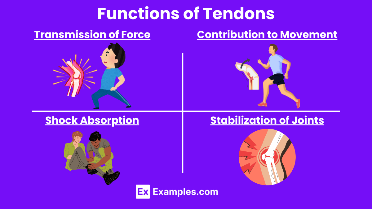
The primary function of tendons is to transmit the force generated by muscles to the bones, facilitating movement. When a muscle contracts, it shortens, pulling on the tendon, which in turn pulls on the bone to which it is attached. This process is crucial for initiating and controlling movement in various parts of the body. For example, when you want to bend your elbow, the biceps muscle contracts, the tendon attached to the biceps pulls on the forearm bone, and your elbow bends.
Tendons play a vital role in enabling both gross and fine motor movements by acting as the connection between muscles and bones. This is evident in tasks ranging from walking and jumping to writing and sewing. The efficiency of tendons in transmitting muscular force with minimal energy loss is key to their functionality. Their ability to stretch slightly under tension and then spring back to their original shape helps maintain movement efficiency and energy conservation.
Tendons also function as shock absorbers, cushioning the impact on joints during high-intensity activities like running or jumping. This shock-absorbing property helps protect muscles and bones from damage caused by sudden, forceful impacts. By absorbing some of the kinetic energy from impacts, tendons reduce the likelihood of injuries such as fractures or muscle strains.
Beyond movement and shock absorption, tendons help stabilize joints, maintaining the alignment and overall posture of the body. This stabilization is crucial during both static poses and dynamic movements, ensuring that joints are not overextended or displaced. The tendons around a joint work together with ligaments and muscles to maintain joint stability and prevent dislocations.
Some tendons, particularly those in the lower limbs like the Achilles tendon, have the ability to store elastic energy when they stretch and then release this energy to aid in movements such as jumping or running. This function is especially important in activities requiring explosive movements, providing a boost that enhances performance and efficiency.
Tendons consist primarily of collagen fibers, specifically type I collagen, which provides high tensile strength and resistance to stretching under tension. These fibers align densely and parallelly, optimizing them for force transmission from muscle to bone. In addition to collagen, tendons include a small proportion of elastin fibers that offer some elasticity, and a matrix of proteoglycans that maintain the structure and hydration of the tendon.
Tendons function as connectors between muscle and bone, facilitating the transmission of forces that enable movement. At the muscular junction, tendons integrate into the muscle tissue through a specialized zone known as the myotendinous junction. Here, muscle fibers taper off and collagen fibers begin, ensuring a secure attachment.
At the bone end, tendons attach through the osteotendinous junction, a highly specialized interface where the tendon’s collagen fibers merge into the bone. This connection strengthens as the tendon fibers gradually transition into fibrocartilage before becoming bone tissue, enhancing the load-bearing capacity and reducing the risk of injury from stress concentration at the interface.
A tendon divides typically into three main parts:
Tendons receive a sophisticated blood supply that originates from multiple sources, enhancing their functionality and health. The primary blood vessels stem from the muscles themselves and from the periosteum at the osteotendinous junctions. Additionally, if present, peritendinous leaflets or a synovial sheath contribute to the vascular network surrounding the tendon.
The arrangement of these vascular networks varies, not only within a single tendon but also among different tendons. Typically, primary vascular trunks create a mesh structure, though sometimes they may form concentric arches or appear irregularly. These networks include small to medium-caliber arteries, which are often accompanied by one or two venous anastomoses.
Within the tendons, three distinct types of microvascular capillary units exist. One type features capillaries running parallel to the tendon’s longitudinal axis before looping back to join a venule. Another type comprises a single arteriole branching into multiple capillary loops, extending in various planes within the tendon. A third type functions as an arterio-venous shunt with short, straight capillaries. This diversity in microvascular structures facilitates efficient diffusion of gases and metabolites across the tendon fibers.
Tendons also possess an intricate lymphatic drainage system, with lymph flowing towards the tendon veins or neighboring venous structures. Importantly, while blood flow to a tendon increases under mechanical stress, lymphatic drainage does not proportionately increase.
Tendon innervation primarily comes from nerve branches linked to the muscle belly and those distributed to the skin. Although innervation is generally sparse, nerves localize mainly to the paratenon, endotenon, and epitenon.
Nerve branches align parallel to the tendon’s central axis and anastomose with others running in transverse and oblique directions. The nerve endings vary, with some connecting to specialized sensory structures like the Golgi tendon organs, and Pacini or Ruffini corpuscles, while others end in free arborizations. The alignment of nerve fibers with the vascular network ensures coordinated responses to physiological demands.
Sympathetic nerves follow the vascular pathways and are crucial for regulating blood flow within the tendon, promoting vasoconstriction and, in certain cases, vasodilation through sensory fibers similar to those in the parasympathetic system or through separate small-diameter sensory fibers.
Tendons can heal naturally but slowly, requiring rest, proper nutrition, and sometimes medical intervention to aid recovery.
The four key symptoms of tendonitis are pain, swelling, tenderness, and reduced movement in the affected area.
Healing tendons involves rest, ice application, compression, elevation, and appropriate physical therapy or exercises.
The four commonly discussed tendons are the Achilles, patellar, rotator cuff, and biceps tendons.
The Achilles tendon is the strongest and toughest tendon, crucial for walking, running, and jumping.

Which of the following best describes a tendon?
A type of muscle
A connective tissue that attaches muscles to bones
A type of bone
A fluid-filled sac
What is the primary function of tendons?
Store calcium
Connect bones to other bones
Transmit force from muscle to bone
Produce red blood cells
Which protein is most abundant in tendons?
Keratin
Hemoglobin
Collagen
Myosin
Tendons are primarily composed of:
Adipose tissue
Epithelial cells
Dense regular connective tissue
Nervous tissue
Which of the following injuries involves a tendon?
Sprain
Fracture
Tendinitis
Contusion
Where is the Achilles tendon located?
Arm
Back
Leg
Neck
Which movement would most likely involve the biceps tendon?
Kicking a ball
Lifting a weight
Running
Jumping
What is the primary difference between tendons and ligaments?
Tendons connect bones to bones, ligaments connect muscles to bones
Tendons connect muscles to bones, ligaments connect bones to bones
Tendons are more flexible than ligaments
Ligaments are more durable than tendons
Which tendon is commonly known as the "patellar tendon"?
Tendon connecting the biceps to the elbow
Tendon connecting the quadriceps to the knee cap
Tendon connecting the deltoid to the shoulder
Tendon connecting the hamstring to the hip
What type of cells are primarily responsible for tendon repair?
Osteocytes
Chondrocytes
Tenocytes
Adipocytes