Preparing for the MCAT requires a thorough understanding of cellular division, a fundamental aspect of the Cellular and Molecular Biology section. Mastery of processes like mitosis and meiosis is essential. This knowledge provides insights into genetic continuity, cell cycle regulation, and variations in genetic makeup, crucial for achieving a high MCAT score.
Learning Objective
In studying "Cellular Division" for the MCAT, you should aim to understand the mechanisms and stages of mitosis and meiosis, including their roles in growth, repair, and reproduction. Analyze how the cell cycle is regulated through checkpoints and the implications of mutations in cell cycle regulators, like p53 and cyclins. Evaluate the differences between somatic cell division and gametogenesis. Additionally, explore the significance of non-disjunction and chromosomal abnormalities in human disease. Apply this knowledge to interpret experimental results and genetic problems in MCAT practice passages, enhancing your preparedness for questions on genetic stability and variability.
Introduction to Cellular Division
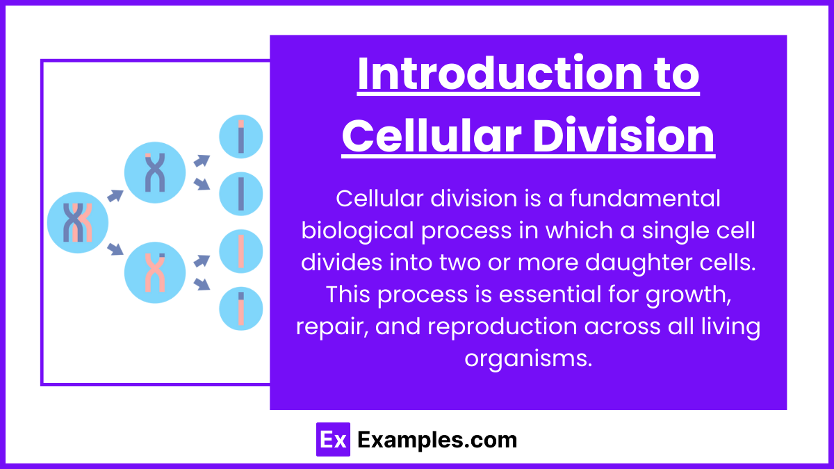
Cellular division is a fundamental biological process in which a single cell divides into two or more daughter cells. This process is essential for growth, repair, and reproduction across all living organisms. Cellular division is categorized primarily into two types: mitosis and meiosis, each playing a distinct role in the life cycle of cells.
Importance of Cellular Division Cellular division supports various biological functions:
Growth and Development: Multicellular organisms rely on mitotic division for growth, allowing an increase in body size and replacement of old cells.
Repair and Regeneration: Tissue damage and wear are addressed through mitosis, which replenishes and maintains tissue integrity.
Reproduction: Meiosis is crucial for sexual reproduction, producing gametes like sperm and eggs that have half the usual number of chromosomes, thereby fostering genetic diversity.
Types of Cellular Division
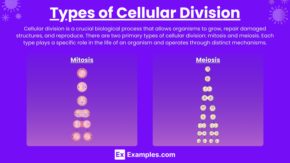
Cellular division is a crucial biological process that allows organisms to grow, repair damaged structures, and reproduce. There are two primary types of cellular division: mitosis and meiosis. Each type plays a specific role in the life of an organism and operates through distinct mechanisms.
Mitosis
Mitosis is the process by which a single cell divides to produce two identical daughter cells. It is fundamental for growth, tissue repair, and asexual reproduction in eukaryotic organisms. Here's how it occurs:
Purpose: Growth, tissue repair, asexual reproduction in eukaryotes.
Process:
Prophase: Chromosomes condense, nuclear envelope breaks down.
Metaphase: Chromosomes align at the cell's equator.
Anaphase: Sister chromatids separate to opposite poles.
Telophase: Nuclear envelopes reform, chromosomes decondense.
Cytokinesis: Cell divides, forming two identical daughter cells.
Meiosis
Meiosis is a specialized form of cellular division that reduces the chromosome number by half, resulting in four haploid cells, each genetically distinct from the parent cell. This process is crucial for sexual reproduction and genetic diversity.
Purpose: Sexual reproduction, genetic diversity.
Process:
Meiosis I: Reduces chromosome number by half; homologous chromosomes separate.
Meiosis II: Similar to mitosis; separates sister chromatids, resulting in four haploid cells.
Chromosomal Abnormalities and Disease Implications
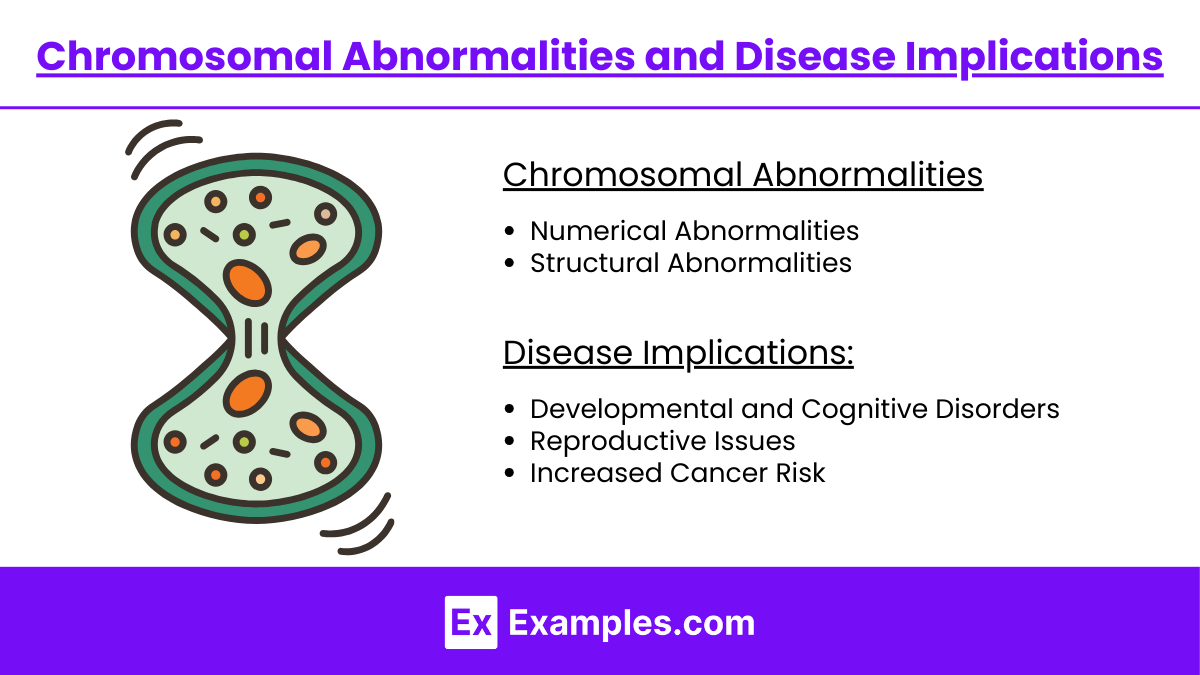
Chromosomal abnormalities involve changes in the number or structure of chromosomes and can lead to a wide range of genetic disorders and health complications. These abnormalities usually occur during cell division, either during mitosis or meiosis, and can be classified into several types, each with different implications for health.
Types of Chromosomal Abnormalities:
Numerical Abnormalities: Such as aneuploidy (e.g., Trisomy 21 leading to Down syndrome) and polyploidy (often resulting in miscarriage).
Structural Abnormalities: Includes deletions (missing chromosome segments), duplications (extra chromosome segments), translocations (segments transferred to other chromosomes), inversions (reversed chromosome segments), and ring chromosomes (ends of a chromosome join to form a ring).
Disease Implications:
Developmental and Cognitive Disorders: Many chromosomal abnormalities affect brain development and function, leading to various degrees of cognitive impairment and physical abnormalities.
Reproductive Issues: Abnormalities like Turner syndrome (monosomy X) or Klinefelter syndrome (XXY) affect sexual development and fertility.
Increased Cancer Risk: Certain genetic changes, particularly translocations and duplications, can disrupt normal cell growth controls and lead to cancer.
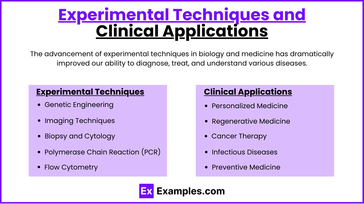
Experimental Techniques and Clinical Applications
The advancement of experimental techniques in biology and medicine has dramatically improved our ability to diagnose, treat, and understand various diseases. Here’s an overview of key experimental techniques and their clinical applications.
Experimental Techniques
Genetic Engineering: Tools like CRISPR-Cas9 allow precise editing of the DNA in genomes, enabling the correction of genetic defects and the study of gene functions.
Imaging Techniques: Advanced imaging, including MRI (Magnetic Resonance Imaging), CT scans (Computed Tomography), and PET scans (Positron Emission Tomography), provide detailed images of the internal structures of the body, aiding in diagnosis and treatment planning.
Biopsy and Cytology: Sampling cells or tissues from the body and examining them under a microscope helps in diagnosing diseases like cancer.
Polymerase Chain Reaction (PCR): This technique amplifies DNA samples, making it easier to analyze and diagnose genetic diseases and infections.
Flow Cytometry: Used to analyze the physical and chemical characteristics of particles in a fluid as they pass through at least one laser. Cell components are fluorescently labeled and then excited by the laser to emit light at varying wavelengths.
Clinical Applications
Personalized Medicine: Genetic and biomarker testing are used to tailor medical treatments to individual genetic profiles, improving efficacy and reducing side effects.
Regenerative Medicine: Stem cell therapy and tissue engineering aim to regenerate damaged tissues and organs. Techniques like 3D bioprinting are now being explored for creating organ structures.
Cancer Therapy: Targeted therapy and immunotherapy, which include techniques such as CAR-T cell therapy, are designed to target cancer cells specifically, which can lead to better outcomes and fewer side effects.
Infectious Diseases: Techniques like PCR and rapid antigen tests have become crucial in the identification and management of infectious diseases, especially with the emergence of pathogens like SARS-CoV-2.
Preventive Medicine: Screening techniques, such as genetic screening and diagnostic imaging, help detect diseases early before symptoms appear, significantly improving treatment success rates.
Examples
Examples 1: Mitosis in Wound Healing
When skin is damaged, cells surrounding the wound undergo mitosis to replace lost or damaged cells. This process exemplifies how mitosis is essential for tissue repair and regeneration, ensuring that new cells are genetically identical to the original cells.
Examples 2: Genetic Variation through Meiosis
During meiosis, processes such as crossing over in prophase I and independent assortment of chromosomes lead to genetic variation in gametes. This variation is crucial for evolution and biodiversity, as it introduces new genetic combinations into populations.
Examples 3: Non-disjunction in Meiosis Leading to Down Syndrome
Non-disjunction, the failure of homologous chromosomes or sister chromatids to separate properly during meiosis, can result in trisomy 21, commonly known as Down Syndrome. This condition occurs when an individual has three copies of chromosome 21.
Examples 4: Cancer Development from Cell Cycle Dysregulation
Cancer can develop when regulatory mechanisms of the cell cycle are disrupted, such as through mutations in genes like p53 or overexpression of cyclins. These mutations can lead to uncontrolled cell division, resulting in tumor growth and progression.
Examples 5: Karyotyping to Identify Chromosomal Abnormalities
Karyotyping is a technique used to detect chromosomal abnormalities, such as deletions, duplications, or translocations, which can be linked to genetic disorders. It involves staining chromosomes to produce a karyotype that allows for the observation of chromosomal differences and anomalies.
Practice Questions:
Question 1
Which phase of mitosis is characterized by the alignment of chromosomes at the center of the cell?
A) Prophase
B) Metaphase
C) Anaphase
D) Telophase
Answer: B) Metaphase
Explanation:
Metaphase is the phase of mitosis where chromosomes align at the metaphase plate, the imaginary line equidistant from the spindle's two poles. This alignment ensures that each daughter cell will receive an equal and exact copy of the chromosomes during the subsequent separation in anaphase.
Question 2
What is a key difference between mitosis and meiosis?
A) Mitosis results in two daughter cells, while meiosis results in four.
B) Mitosis involves one division cycle, whereas meiosis includes two.
C) Only meiosis involves chromosomes aligning at the cell's equator.
D) Only mitosis contributes to genetic diversity.
Answer: A) Mitosis results in two daughter cells, while meiosis results in four.
Explanation:
The primary distinction between mitosis and meiosis is that mitosis results in the production of two genetically identical diploid cells, whereas meiosis produces four genetically distinct haploid cells. This difference is fundamental to the respective roles of these processes: growth and repair in mitosis versus reproduction in meiosis.
Question 3
Which event occurs during anaphase of mitosis?
A) Chromosomes condense and become visible.
B) The nuclear envelope re-forms around separated chromosomes.
C) Sister chromatids separate and move to opposite poles.
D) Crossing over between homologous chromosomes.
Answer: C) Sister chromatids separate and move to opposite poles.
Explanation:
During anaphase of mitosis, the sister chromatids, previously paired during metaphase, are pulled apart by the spindle fibers. Each chromatid, now an individual chromosome, moves to opposite poles of the cell. This ensures that each new daughter cell will contain the same number of chromosomes as the original cell. Anaphase ensures the correct distribution of chromosomes, critical for cellular function and genetic integrity.

