
Cytoskeleton

Preparing for the MCAT demands a thorough understanding of the cytoskeleton, a fundamental component of the Cellular and Molecular Biology foundation. Mastery of its structure, functions, and dynamics is essential. This knowledge elucidates cell shape, movement, and intracellular transport, crucial for achieving a high MCAT score.
Learning Objective
In studying “Cytoskeleton” for the MCAT, you should aim to understand the composition and functions of microtubules, microfilaments, and intermediate filaments. Analyze how these structures support cellular shape, enable intracellular transport, and facilitate cellular movements. Evaluate the mechanisms of cytoskeletal dynamics during cell division and motility. Additionally, explore the role of the cytoskeleton in signal transduction and its implications in diseases such as cancer. Apply this knowledge to interpret experimental results and solve complex biological problems in MCAT practice passages, enhancing your understanding of cellular behavior in physiological and pathological conditions.
Overview of Cytoskeletal
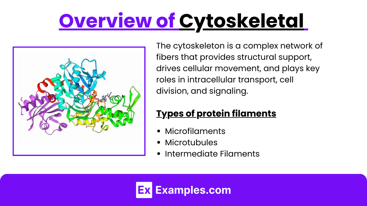
The cytoskeleton is a complex network of fibers that provides structural support, drives cellular movement, and plays key roles in intracellular transport, cell division, and signaling. It consists of three main types of protein filaments:
1. Microfilaments
- Main Component: Actin, a protein that forms thin, flexible fibers.
- Functions:
- Shape and support the cell.
- Enable the cell to move and divide.
- Important in muscle contraction.
2. Microtubules
- Main Component: Tubulin, which forms rigid, hollow tubes.
- Functions:
- Support the cell and give it shape.
- Guide organelle movement within the cell.
- Help pull chromosomes apart during cell division.
- Form structures like cilia and flagella for cell movement.
3. Intermediate Filaments
- Main Component: Various proteins like vimentin and keratin, depending on the cell type.
- Functions:
- Strengthen the cell against mechanical stress.
- Help hold organelles in place.
- Support the nucleus and help keep its shape.
Cytoskeletal Functions in Cell Division and Motility
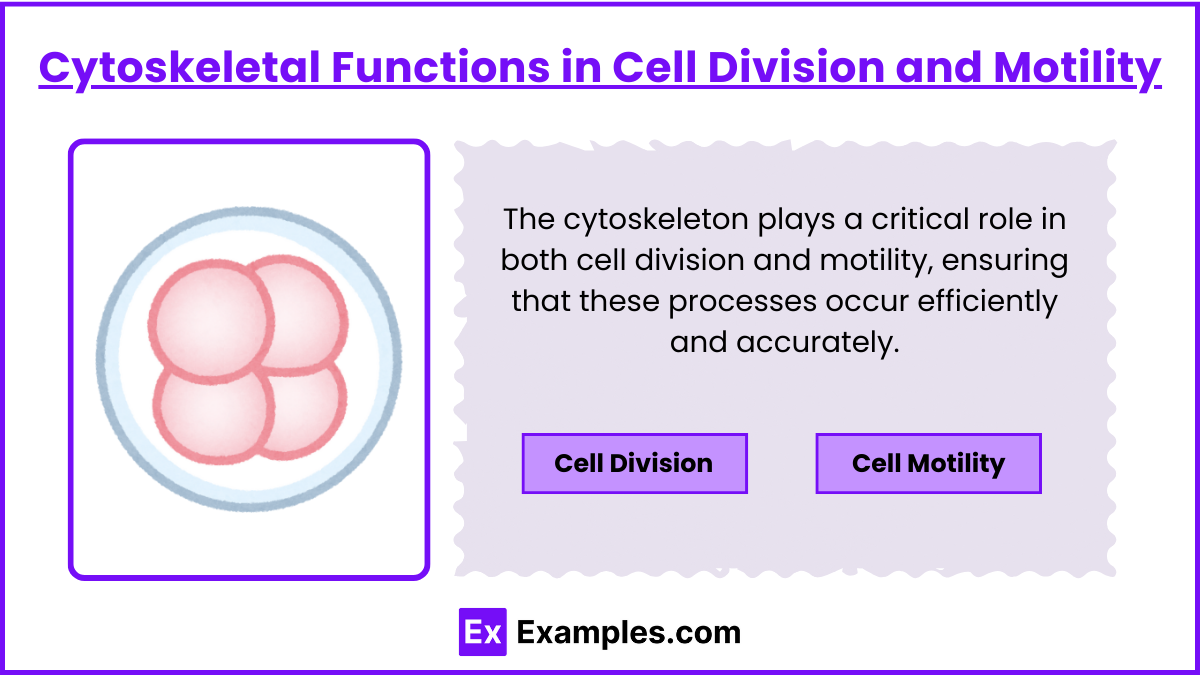
The cytoskeleton plays a critical role in both cell division and motility, ensuring that these processes occur efficiently and accurately. Below is an overview of how the cytoskeleton supports these functions:
1. Cell Division
- Microtubules:
- Form the mitotic spindle, which pulls chromosomes apart during division.
- Attach to chromosomes via kinetochores, ensuring proper separation to daughter cells.
- Actin Filaments:
- Form the contractile ring during cytokinesis to split the cell in two.
- Actin and myosin work together to constrict and divide the cell membrane.
2. Cell Motility
- Actin Filaments:
- Drive cell crawling by extending cell projections (lamellipodia and filopodia).
- Actin polymerization pushes the cell forward while contraction pulls the cell body.
- Microtubules:
- Stabilize the cell’s leading edge and guide its directional movement.
- Transport essential molecules and align internal components for coordination.
- Intermediate Filaments:
- Provide structural support, maintaining cell shape during movement.
Pathological Implications of Cytoskeletal Dysfunctions
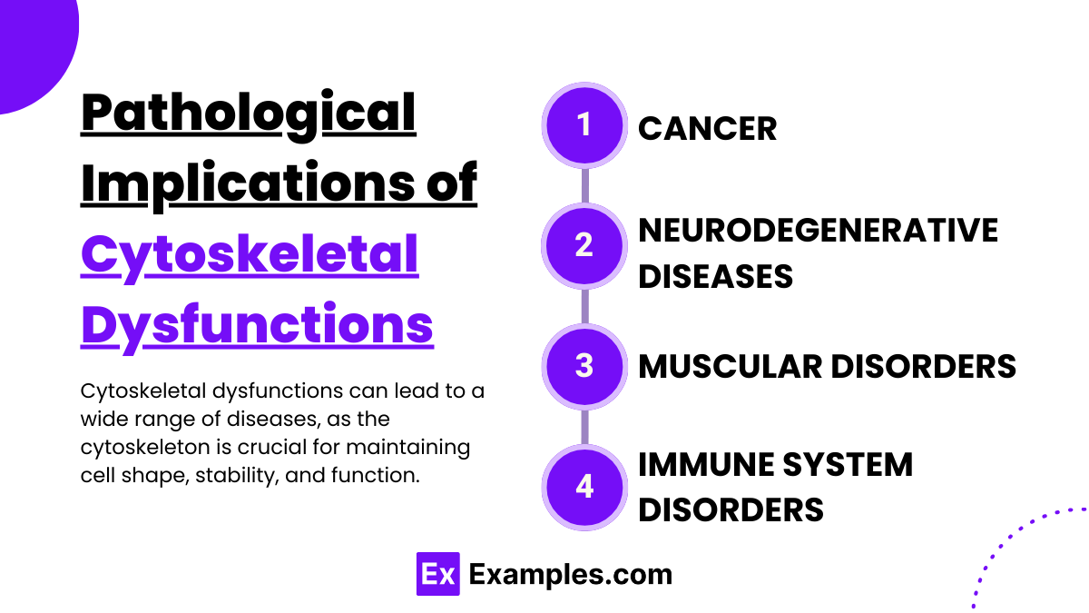
Cytoskeletal dysfunctions can lead to a wide range of diseases, as the cytoskeleton is crucial for maintaining cell shape, stability, and function. Below are key examples of how cytoskeletal abnormalities contribute to pathology:
1. Cancer
- Cell Migration and Metastasis:
- Abnormalities in actin filaments and microtubules can enhance cell movement, contributing to the spread of cancer cells (metastasis).
- Altered signaling pathways affecting cytoskeletal dynamics lead to uncontrolled cell migration and invasion into other tissues.
2. Neurodegenerative Diseases
- Axonal Transport Defects:
- Microtubule dysfunctions disrupt the transport of essential materials (proteins, organelles) along nerve cell axons, contributing to diseases like Alzheimer’s, Parkinson’s, and ALS (Amyotrophic Lateral Sclerosis).
- Tau protein abnormalities cause microtubule instability, leading to neuron degeneration.
3. Muscular Disorders
- Muscle Weakness and Dystrophies:
- Mutations in actin or associated proteins (e.g., dystrophin) impair muscle fiber integrity and function, leading to conditions like muscular dystrophy.
- Cytoskeletal damage affects muscle cell contraction and repair.
4. Immune System Disorders
- Impaired Immune Response:
- Cytoskeletal dysfunctions in immune cells (e.g., actin filament defects) hinder the movement and activity of white blood cells, affecting the body’s ability to respond to infections or inflammation.
Examples
Examples 1: Microtubule Organization in Neurons
- Microtubules provide structural support and facilitate axonal transport, which is essential for the proper functioning of neurons. Disruptions in microtubule stability can lead to neurological disorders such as Alzheimer’s disease.
Examples 2: Actin Filament Role in Muscle Contraction
- Actin filaments, in conjunction with myosin, are crucial for muscle contraction. This interaction is fundamental to understanding how muscles contract and relax, and disruptions can lead to muscular diseases.
Examples 3: Intermediate Filaments in Epithelial Tissue Integrity
- Intermediate filaments provide mechanical support for epithelial cells that form barriers, such as the skin. Mutations in keratin, a type of intermediate filament, can cause skin blistering diseases such as epidermolysis bullosa simplex.
Examples 4: Microtubules in Mitotic Spindle Formation
- During mitosis, microtubules organize into a spindle apparatus that separates chromosomes into daughter cells. This process is critical for cell division and is a target for anticancer drugs that disrupt microtubule dynamics.
Examples 5: Actin Dynamics in Cell Motility
- Actin polymerization at the leading edge of a cell drives the formation of structures like lamellipodia and filopodia, enabling cell movement. This is vital in processes such as wound healing and cancer cell metastasis.
Practice Questions:
Question 1
Which cytoskeletal component is primarily responsible for the contraction of muscle cells?
A) Microtubules
B) Microfilaments
C) Intermediate filaments
D) Motor proteins
Answer: B) Microfilaments
Explanation:
Microfilaments, particularly those composed of actin, are crucial for muscle contraction. In muscle cells, actin filaments interact with myosin (a type of motor protein), allowing for the sliding filament mechanism that causes muscle contraction. This is essential knowledge for understanding muscular movement and dynamics.
Question 2
During cell division, which structure is responsible for pulling chromosomes apart to opposite poles of the cell?
A) Centrosome
B) Golgi apparatus
C) Mitotic spindle
D) Nuclear envelope
Answer: C) Mitotic spindle
Explanation:
The mitotic spindle, composed of microtubules, is crucial during mitosis for chromosome alignment and separation. Microtubules emanating from opposite spindle poles attach to the chromosomes’ centromeres via kinetochore complexes and help in their segregation to daughter cells, ensuring proper division and genetic stability.
Question 3
Which type of cytoskeletal filament is primarily involved in maintaining the strength and structural integrity of skin cells?
A) Actin filaments
B) Microtubules
C) Intermediate filaments
D) Elastic fibers
Answer: C) Intermediate filaments
Explanation:
Intermediate filaments, particularly keratins in epithelial cells, provide mechanical strength and resilience to cells against physical stress. In skin cells, keratin intermediate filaments are essential for maintaining cell structure and preventing tearing or blistering under mechanical stress, demonstrating their critical role in tissue integrity.