Preparing for the MCAT requires a thorough understanding of embryology, a key component of the Organ Systems foundation. Mastery of embryonic development stages, cellular differentiation, and organogenesis is essential. This knowledge provides insights into congenital anomalies, developmental biology, and prenatal health, crucial for achieving a high MCAT score.
Learning Objective
In studying "Embryology" for the MCAT, you should aim to understand the fundamental processes of early human development from fertilization through to fetal growth. Analyze the stages of embryogenesis including zygote formation, blastulation, gastrulation, and neurulation. Evaluate the mechanisms of cellular differentiation and tissue specialization. Additionally, explore the impact of genetic and environmental factors on embryonic development and examine common developmental disorders. Apply this knowledge to interpret clinical scenarios and experimental results in MCAT practice passages, enhancing your understanding of developmental biology and its implications for human health and disease.
Overview of Embryology
Embryology is the branch of biology that studies the prenatal development of gametes (sex cells), fertilization, and development of embryos and fetuses. Additionally, embryology encompasses the study of congenital disorders that occur before birth. Here's a concise overview of the fundamental stages and processes in embryological development:
Gametogenesis: The production of gametes (sperm in males via spermatogenesis and eggs in females via oogenesis).
Fertilization: The fusion of an egg and sperm cell that forms a zygote, initiating the development of a new individual.
Cleavage and Blastulation:
Rapid mitotic divisions of the zygote called cleavage.
Formation of the blastocyst, a structure consisting of an inner cell mass (future embryo) and an outer layer (future placenta).
Gastrulation: A reorganization process where the blastocyst forms a three-layered structure with distinct germ layers:
Ectoderm: Develops into the nervous system and skin.
Mesoderm: Forms muscles, bones, and the cardiovascular system.
Endoderm: Creates the digestive tract and other internal organs.
Organogenesis: The development of organs from the three germ layers, including the formation of the neural tube from the ectoderm, which becomes the brain and spinal cord.
Neurulation: The formation and closure of the neural tube, which is crucial for proper brain and spinal cord development. Failures in this process can lead to neural tube defects like spina bifida.
Fertilization and Early Development in Embryology
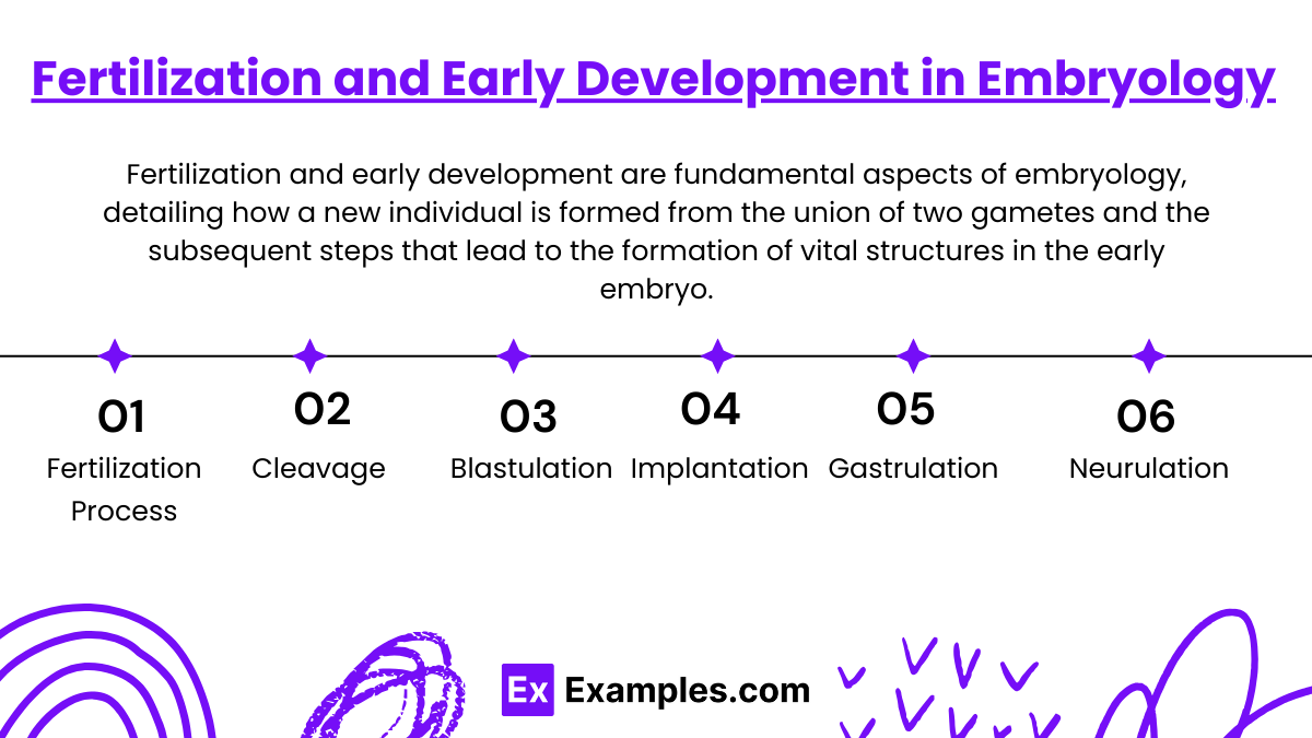
Fertilization and early development are fundamental aspects of embryology, detailing how a new individual is formed from the union of two gametes and the subsequent steps that lead to the formation of vital structures in the early embryo. Here’s a concise overview of these critical stages:
Fertilization Process
Fertilization occurs when a sperm penetrates an egg, leading to the formation of a zygote.
This event restores the diploid chromosome number and activates egg metabolism, initiating embryonic development.
Cleavage
The zygote undergoes rapid mitotic divisions known as cleavage, producing smaller cells called blastomeres.
These cells form a morula, a solid ball of cells.
Blastulation
The morula develops into a blastocyst, which features a fluid-filled cavity (blastocoel) and two distinct components:
Inner Cell Mass (ICM): Develops into the embryo.
Trophoblast Layer: Contributes to the placenta and initiates implantation into the uterine wall.
Implantation
The blastocyst attaches to the uterine wall, where it begins to implant.
The trophoblast layer differentiates and interacts with the endometrium to form the embryonic part of the placenta.
Gastrulation
Cells of the inner cell mass reorganize into three primary germ layers:
Ectoderm: Becomes the nervous system and skin.
Mesoderm: Forms muscles, bones, and the circulatory system.
Endoderm: Develops into the internal organs like the digestive tract and lungs.
Neurulation
The neural plate forms from the ectoderm, folds to create the neural tube, the precursor to the central nervous system.
Neurulation and Organogenesis in Embryology
Neurulation and organogenesis are critical phases in embryonic development, during which the neural structures and major organ systems begin to form. Understanding these processes provides insights into both normal developmental biology and the origins of various congenital anomalies.
Neurulation
Neurulation is the process that leads to the formation of the central nervous system (CNS) and marks the beginning of the body's complex neural structure development.
Formation of the Neural Plate: Early in development, a portion of the ectoderm on the dorsal surface of the embryo thickens to form the neural plate.
Formation of the Neural Tube: As neurulation progresses, the edges of the neural plate begin to rise and fold towards each other, eventually fusing to form the neural tube. This tube will differentiate into the brain and spinal cord.
Closure of the Neural Tube: The neural tube closes starting from the middle and extending towards both ends. Incomplete closure at either end can result in neural tube defects such as spina bifida (lower end) or anencephaly (upper end).
Organogenesis
Organogenesis is the phase in which the embryonic germ layers differentiate into the various organs and tissues of the body.
Ectoderm: Besides contributing to the neural tube, the ectoderm also forms the epidermis, hair, nails, and parts of the eye and inner ear.
Mesoderm: This layer develops into a diverse range of structures, including the skeletal system, muscular system, cardiovascular system, kidneys, and much of the reproductive system. The mesoderm also gives rise to connective tissues throughout the body.
Endoderm: The innermost layer forms the lining of the digestive and respiratory tracts and develops into associated organs such as the liver, pancreas, and thyroid gland.
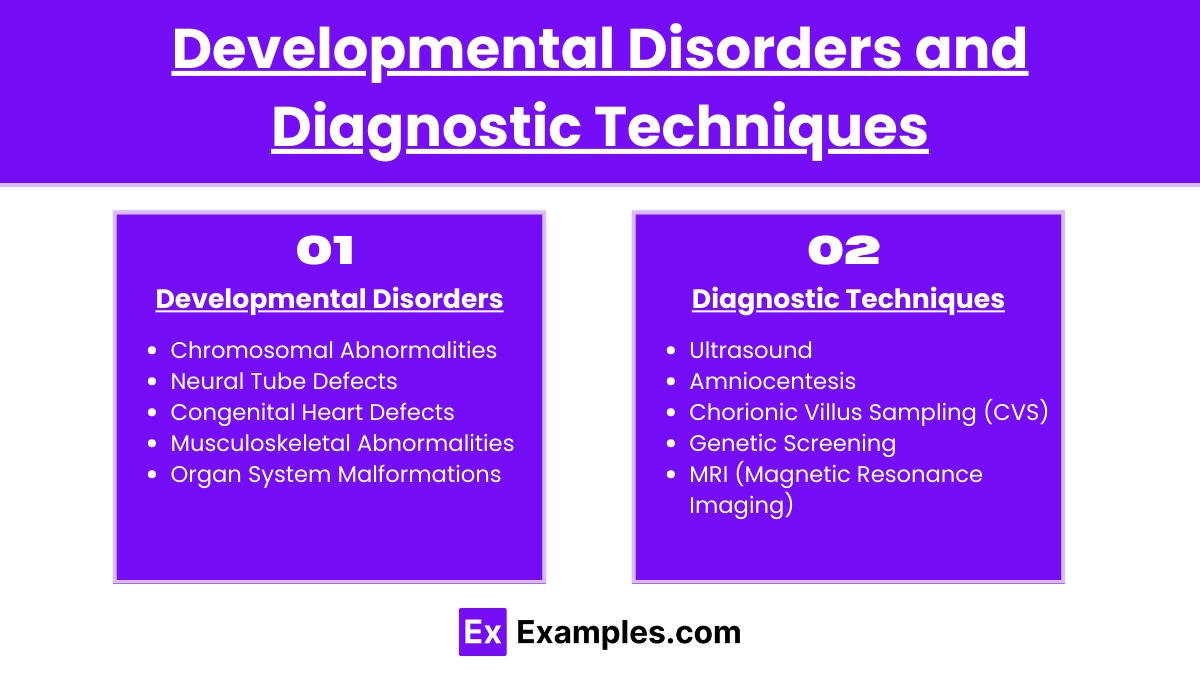
Developmental Disorders and Diagnostic Techniques
Developmental disorders arise from disruptions in normal embryological growth and differentiation. These conditions can affect various systems and have lifelong impacts on an individual's health and functioning. Understanding these disorders and the diagnostic techniques used to identify them is crucial for early intervention and management.
Types of Developmental Disorders:
Chromosomal Abnormalities: Disorders like Down syndrome, Turner syndrome, and Klinefelter syndrome result from anomalies in chromosome number or structure.
Neural Tube Defects: Conditions such as spina bifida and anencephaly occur due to improper closure of the neural tube during early development.
Congenital Heart Defects: Structural issues in the heart present from birth, such as septal defects or congenital valve disorders.
Musculoskeletal Abnormalities: This category includes limb malformations and congenital hip dysplasia.
Organ System Malformations: Can affect any body system, such as the digestive or respiratory systems, leading to conditions like congenital diaphragmatic hernia.
Diagnostic Techniques:
Early and accurate diagnosis of developmental disorders is key to managing health outcomes and planning interventions.
Ultrasound: Routinely used during pregnancy to assess fetal growth and development. High-resolution ultrasounds can detect structural abnormalities in the fetus.
Amniocentesis: Involves sampling amniotic fluid around the fetus to detect chromosomal abnormalities and genetic disorders. Typically performed around the 15th week of pregnancy.
Chorionic Villus Sampling (CVS): A prenatal test that involves taking a sample of tissue from the placenta to test for genetic abnormalities. CVS can be performed earlier than amniocentesis, usually between the 10th and 12th weeks of pregnancy.
Genetic Screening: Tests such as maternal blood screening can detect markers that suggest the risk of chromosomal conditions like Down syndrome.
MRI (Magnetic Resonance Imaging): Provides detailed imaging that can help diagnose complex brain and spinal cord anomalies and other internal structural defects.
Examples
Examples 1: Zygote to Blastocyst Transformation
Following fertilization, the zygote undergoes several divisions, transitioning through stages such as morula and blastocyst. The blastocyst's inner cell mass will eventually form the embryo, while the outer cell layer, the trophoblast, develops into part of the placenta.
Examples 2: Gastrulation Process
Gastrulation is a pivotal event in early development where the blastula is reorganized into a multilayered structure forming the three primary germ layers: ectoderm, mesoderm, and endoderm. This process sets the stage for all future organ and tissue development.
Examples 3: Neural Tube Formation (Neurulation)
During neurulation, the cells in the ectoderm layer form the neural tube, which is the precursor to the central nervous system, comprising the brain and spinal cord. Defects in this process can lead to serious conditions such as spina bifida.
Examples 4: Impact of Teratogens
Exposure to teratogens like alcohol or certain medications during pregnancy can disrupt normal embryonic development, leading to fetal alcohol syndrome or limb malformations. These examples highlight the critical influence of environmental factors on embryonic health.
Examples 5: Diagnostic Technique: Ultrasound Imaging
Ultrasound is routinely used during pregnancy to monitor the developmental progress of the fetus, detect congenital anomalies, and evaluate fetal health. This non-invasive technique allows for real-time observation of the fetus within the womb.
Practice Questions:
Question 1
What structure forms from the ectoderm during embryonic development?
A) Heart
B) Spinal cord
C) Lungs
D) Liver
Answer: B) Spinal cord
Explanation:
The spinal cord, along with the brain, is formed from the neural tube, which originates from the ectoderm layer. This layer primarily gives rise to structures associated with the nervous system and skin, making B the correct answer. The other options, heart (mesoderm), lungs (endoderm), and liver (endoderm), develop from different germ layers.
Question 2
During which embryonic process do the three primary germ layers form?
A) Fertilization
B) Gastrulation
C) Organogenesis
D) Cleavage
Answer: B) Gastrulation
Explanation:
Gastrulation is the critical phase in embryonic development where the single-layered blastula is reorganized into a trilaminar structure forming the ectoderm, mesoderm, and endoderm. These layers are foundational for all future organ and tissue development, making Gastrulation (B) the correct answer. The other stages listed do not involve germ layer formation.
Question 3
Which condition results from improper closure of the neural tube during development?
A) Cleft palate
B) Down syndrome
C) Spina bifida
D) Congenital heart defect
Answer: C) Spina bifida
Explanation:
Spina bifida is a congenital defect that occurs when the neural tube does not close completely. This results in an incomplete development of the spinal cord and vertebrae. It is a direct result of a failure in neurulation, making C the correct answer. The other conditions listed are not directly related to neurulation errors but involve other developmental processes or genetic issues.

