
Muscular System

Preparing for the MCAT requires a thorough understanding of the muscular system, essential for comprehending how muscles function and interact within various organ systems. Studying key concepts such as muscle contraction, types of muscle tissue, and their roles in movement and stability is vital. Mastering these concepts will bolster your biological systems knowledge, crucial for a high MCAT score
Learning Objective
In studying the "Muscular System" for the MCAT, you should aim to understand the structure and function of different muscle types, including skeletal, cardiac, and smooth muscles. Explore the biochemical pathways of muscle contraction and energy metabolism. Examine the roles of actin and myosin in muscle movement, and how neural input regulates muscle activity. Additionally, understand the implications of muscular system disorders and their treatments. Apply this knowledge to analyze physiological data and clinical scenarios in MCAT practice passages, enhancing your ability to interpret how muscular diseases and treatments affect overall body function.
Introduction to the Muscular System
The muscular system is a complex network of tissues that play a crucial role in the overall functioning of the human body. It enables movement, supports posture, and is vital in circulatory and respiratory systems. Here’s an introductory overview:
There are three main types of muscles in the human body:
Skeletal Muscles: These are attached to bones and are responsible for voluntary movements. They are controlled by the nervous system, and their activation helps in activities like running, lifting, and all forms of physical activity.
Smooth Muscles: Located within the walls of internal organs such as the stomach, intestines, and blood vessels, these muscles control involuntary movements. They help in functions like digestion, circulation, and regulation of blood pressure.
Cardiac Muscle: This type of muscle is found only in the heart. It is highly specialized and designed to continuously pump blood throughout life without fatigue. The contractions of the cardiac muscle are also involuntary.
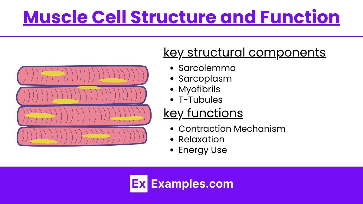
Muscle Cell Structure and Function
Muscle cells, also known as muscle fibers, are specialized cells that are key components of the muscular system. Their structure is uniquely designed to facilitate contraction, which is essential for all types of muscle movements. Here’s a detailed look at the structure and function of muscle cells:
Structure of Muscle Cells
Muscle cells, also known as muscle fibers, have a unique structure optimized for contraction and movement. Here’s a simplified overview of their key structural components:
Sarcolemma: The cell membrane that regulates entry and exit of substances.
Sarcoplasm: Cytoplasm containing energy reserves like glycogen and oxygen-binding myoglobin.
Myofibrils: Rod-like structures filled with sarcomeres, the basic functional units of muscle fibers, consisting of actin (thin filaments) and myosin (thick filaments).
T-Tubules: Extensions of the sarcolemma that help transmit electrical signals deep into the cell.
Sarcoplasmic Reticulum (SR): Stores and releases calcium ions, vital for muscle contraction.
Function of Muscle Cells
Muscle cells, or muscle fibers, have several key functions related to movement and stability of the body. Here’s a streamlined explanation of their primary roles:
Contraction Mechanism: Triggered by electrical impulses that lead to calcium release from the SR, enabling myosin heads to bind to actin and shorten the sarcomere.
Relaxation: Occurs when calcium ions are pumped back into the SR, allowing the muscle to lengthen and relax.
Energy Use: ATP is used by myosin for contraction cycles and detachment from actin.
Biochemical Pathways of Muscle Contraction
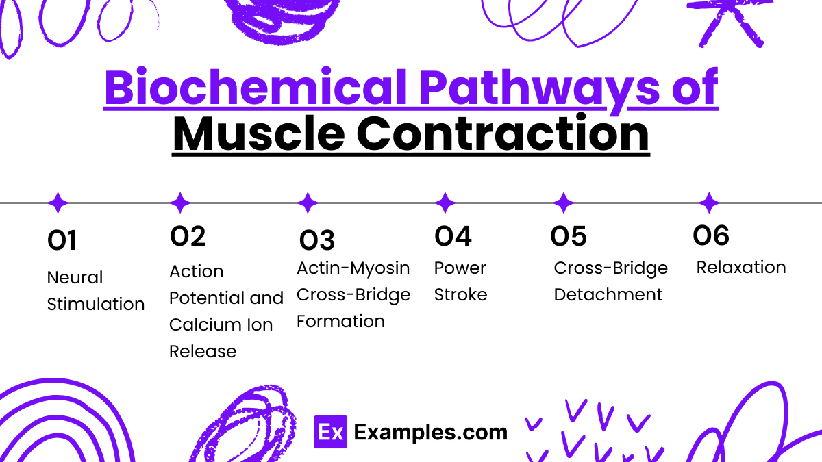
Muscle contraction involves a series of biochemical events within the muscle cell that are crucial for movement. Here’s a detailed explanation of the biochemical pathways involved in this process:
1. Neural Stimulation
Neurotransmitter Release: Muscle contraction begins when a motor neuron releases the neurotransmitter acetylcholine (ACh) at the neuromuscular junction.
ACh Binding: ACh binds to receptors on the sarcolemma (muscle cell membrane), triggering an action potential that spreads along the muscle fiber.
2. Action Potential and Calcium Ion Release
Action Potential Propagation: The action potential travels along the sarcolemma and down the T-tubules, deep into the muscle fiber.
Calcium Release: This electrical signal triggers the sarcoplasmic reticulum (SR) to release stored calcium ions into the sarcoplasm.
3. Actin-Myosin Cross-Bridge Formation
Troponin and Tropomyosin Regulation: Calcium ions bind to troponin, a regulatory protein on the thin actin filaments. This binding causes troponin to change shape, moving tropomyosin (another regulatory protein) away from actin’s myosin-binding sites.
Cross-Bridge Attachment: Myosin heads, which are part of the thick filaments, attach to these newly exposed binding sites on actin, forming cross-bridges.
4. Power Stroke
ADP and Pi Release: Attached myosin heads pivot, pulling the actin filament toward the center of the sarcomere in a movement known as the power stroke. This movement shortens the muscle fiber, causing contraction. ADP and inorganic phosphate (Pi), previously bound to myosin, are released during this step.
5. Cross-Bridge Detachment
ATP Binding: A new molecule of ATP binds to the myosin head, causing it to detach from actin.
ATP Hydrolysis: The myosin head hydrolyzes ATP to ADP and Pi, using the energy to return to its original "cocked" position.
6. Relaxation
Calcium Reuptake: When neural stimulation ceases, calcium ions are pumped back into the SR, lowering the cytoplasmic calcium concentration.
Troponin and Tropomyosin Reset: As calcium levels fall, troponin returns to its original shape, allowing tropomyosin to cover actin’s myosin-binding sites again. This prevents further cross-bridge formation.
Muscle Fiber Lengthens: Without cross-bridges, the muscle fiber relaxes and lengthens.
Muscular System Disorders
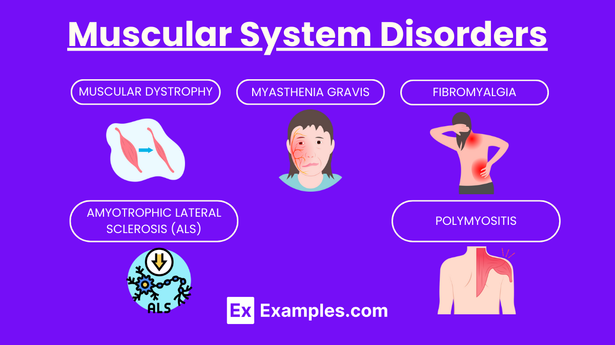
Disorders of the muscular system can impact the body's ability to move, maintain posture, and perform vital functions. Here are some common muscular system disorders explained simply:
1. Muscular Dystrophy
What It Is: A group of inherited disorders that cause muscle weakness and loss of muscle mass over time.
Symptoms: Progressive muscle weakness, difficulty walking, and loss of mobility.
Cause: Genetic mutations affect the production of proteins needed to build and maintain healthy muscles.
2. Myasthenia Gravis
What It Is: An autoimmune disorder where the immune system mistakenly attacks the connections between nerves and muscles.
Symptoms: Muscle weakness that worsens with activity and improves with rest, drooping eyelids, difficulty swallowing and speaking.
Cause: Antibodies block or destroy muscle receptor sites, preventing muscle contraction.
3. Amyotrophic Lateral Sclerosis (ALS)
What It Is: Also known as Lou Gehrig’s disease, ALS is a progressive neurodegenerative disease that affects nerve cells in the brain and spinal cord, leading to loss of muscle control.
Symptoms: Weakness in the limbs, slurred speech, difficulty breathing and swallowing.
Cause: While the exact cause is unknown, it involves the degeneration and death of motor neurons.
4. Fibromyalgia
What It Is: A condition characterized by widespread musculoskeletal pain accompanied by fatigue, sleep, memory, and mood issues.
Symptoms: Widespread pain, fatigue, sleep disturbances, and cognitive difficulties.
Cause: Unknown, but it can be linked to genetics, infections, physical or emotional trauma.
5. Polymyositis
What It Is: An inflammatory muscle disease that causes significant weakness, primarily affecting the muscles closest to the trunk.
Symptoms: Muscle weakness, difficulty climbing stairs or lifting arms, problems with swallowing or breathing.
Cause: The immune system mistakenly attacks muscle tissues, though the exact trigger is unclear.
Examples
Example 1:Muscle Contraction
A sprinter starts a race.
During the sprint, the sprinter’s leg muscles repeatedly perform rapid and powerful contractions. This involves the sliding filament mechanism where actin and myosin fibers slide over each other, shortening the muscle and generating movement.
Example 2:Energy Metabolism in Muscles
Running a marathon.
Over the course of a marathon, muscle cells metabolize stored glycogen through glycolysis and oxidative phosphorylation to continuously supply ATP needed for sustained muscle contraction, shifting from anaerobic to aerobic metabolism as the race progresses.
Example 3:Muscular Dystrophy
Progressive muscle weakness.
In muscular dystrophy, genetic mutations lead to the deterioration of muscle fibers, resulting in progressive strength loss. This condition illustrates the importance of dystrophin, a protein essential for muscle fiber integrity.
Example 4:Neuromuscular Junction Disorder
Myasthenia gravis.
Myasthenia gravis is an autoimmune disorder where antibodies block or destroy nicotinic acetylcholine receptors at the neuromuscular junction. This disruption leads to muscle weakness and fatigue, particularly evident during repetitive muscle use.
Example 5:Calcium's Role in Muscle Contraction
Lifting a heavy object.
When lifting, calcium ions are released from the sarcoplasmic reticulum into the muscle cytoplasm, binding to troponin. This changes the shape of tropomyosin on the actin filaments, exposing binding sites for myosin and initiating contraction.
Practice Questions
Question 1:
What role do calcium ions play in muscle contraction?
A) They break down ATP to release energy.
B) They bind to myosin, allowing it to attach to actin.
C) They trigger the release of acetylcholine at neuromuscular junctions.
D) They bind to troponin, causing a conformational change that exposes binding sites on actin.
Answer: D) They bind to troponin, causing a conformational change that exposes binding sites on actin.
Explanation:
Calcium ions are crucial in muscle contraction because they bind to troponin, which is part of the thin filament in muscle fibers. This binding causes troponin to change shape, moving tropomyosin away from the active sites on actin filaments, thereby allowing myosin heads to bind to these sites and initiate contraction. This process is essential for the sliding filament mechanism of muscle contraction.
Question 2:
Which type of muscle tissue is striated and under involuntary control?
A) Skeletal muscle
B) Smooth muscle
C) Cardiac muscle
D) Epithelial tissue
Answer: C) Cardiac muscle
Explanation:
Cardiac muscle tissue is unique in that it is striated like skeletal muscle, allowing for forceful contractions, but it operates under involuntary control, similar to smooth muscle. This combination is essential for the continuous and rhythmic contractions necessary for heart function, independent of conscious control.
Question 3:
During a marathon, which metabolic pathway is primarily used by muscle cells to generate ATP after the first hour?
A) Phosphagen system
B) Anaerobic glycolysis
C) Aerobic respiration
D) Protein catabolism
Answer: C) Aerobic respiration
Explanation:
During prolonged activities like a marathon, after the initial stores of ATP and creatine phosphate are depleted, and once the limited capacity of anaerobic glycolysis is surpassed, muscle cells rely predominantly on aerobic respiration. This pathway uses oxygen to metabolize carbohydrates, fats, and sometimes proteins to produce ATP, and it is the most efficient way to generate large amounts of ATP necessary for endurance activities.