
Neural cells

Preparing for the MCAT demands a deep understanding of neural cells, fundamental to the Organ Systems foundation. Key concepts include neuron structure, synaptic function, and neural communication. Proficiency in these topics provides crucial insights into how neural networks operate, aiding in the mastery of complex biological interactions necessary for a high MCAT score.
Learning Objective
In studying "Neural Cells" for the MCAT, you should aim to understand the structure, function, and types of neural cells, including neurons and glial cells. Explore the mechanisms of action potentials, synaptic transmission, and neurotransmitter functions. Delve into how neural circuits form and the role of neuroplasticity in learning and memory. Additionally, assess how neural cell dysfunction contributes to neurological disorders. Apply this knowledge to interpret neurological phenomena, analyze clinical implications, and solve practice questions on the MCAT, focusing on how neural cells underpin both normal and pathological conditions in the nervous system.
Understanding Neural Cell Structure and Function
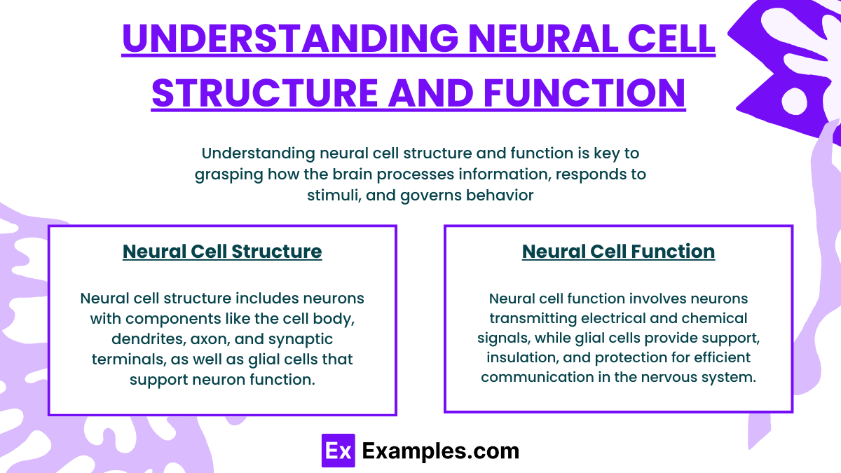
Understanding neural cell structure and function is key to grasping how the brain processes information, responds to stimuli, and governs behavior. Here’s an essential guide to neural cells, covering both neurons and glial cells:
Neuron Structure
Neurons are specialized cells designed to transmit information throughout the body. Their structure is uniquely suited to their role in communication:
Cell Body (Soma): The main part of a neuron, containing the nucleus and most of the cell's organelles. It integrates signals received from dendrites and generates outgoing signals.
Dendrites: These are branched extensions from the soma that receive signals from other neurons. The larger the number of dendrites, the more signals a neuron can receive.
Axon: A long, slender projection that transmits electrical impulses away from the cell body toward other neurons or muscles. Some axons are covered with a myelin sheath, which speeds up signal transmission.
Axon Terminals (Synaptic Terminals): The endpoints of an axon where neurotransmitters are stored and released to communicate with neighboring neurons through the synapse.
Synapse: The junction between two neurons, consisting of a presynaptic ending that contains neurotransmitters, the synaptic cleft, and the postsynaptic ending with receptor sites.
Neuron Function
The primary function of neurons is to receive, process, and transmit information through both electrical and chemical signals:
Electrical Signaling (Action Potentials): Neurons transmit information via electrical impulses known as action potentials. These occur when the neuron's membrane potential becomes more positive, triggering a down-the-line reaction along the axon.
Chemical Signaling (Synaptic Transmission): At the synapse, the electrical signal triggers the release of neurotransmitters into the synaptic cleft. These chemicals bind to receptors on the receiving neuron, continuing the signal.
Glial Cell Structure and Function
Glial cells, or glia, are non-neuronal cells that provide support and protection for neurons in the central and peripheral nervous systems. They are crucial for neural function:
Astrocytes: Regulate blood flow to neurons, maintain the chemical environment, and participate in neurotransmitter regulation at the synaptic cleft.
Oligodendrocytes (CNS) and Schwann Cells (PNS): These cells create the myelin sheath around axons, which is essential for fast signal transmission.
Microglia: Act as the primary immune defense in the central nervous system, removing waste and debris from damaged cells.
Ependymal Cells: Line the spinal cord and ventricles of the brain, producing cerebrospinal fluid that cushions the CNS.
Mechanisms of Neural Communication
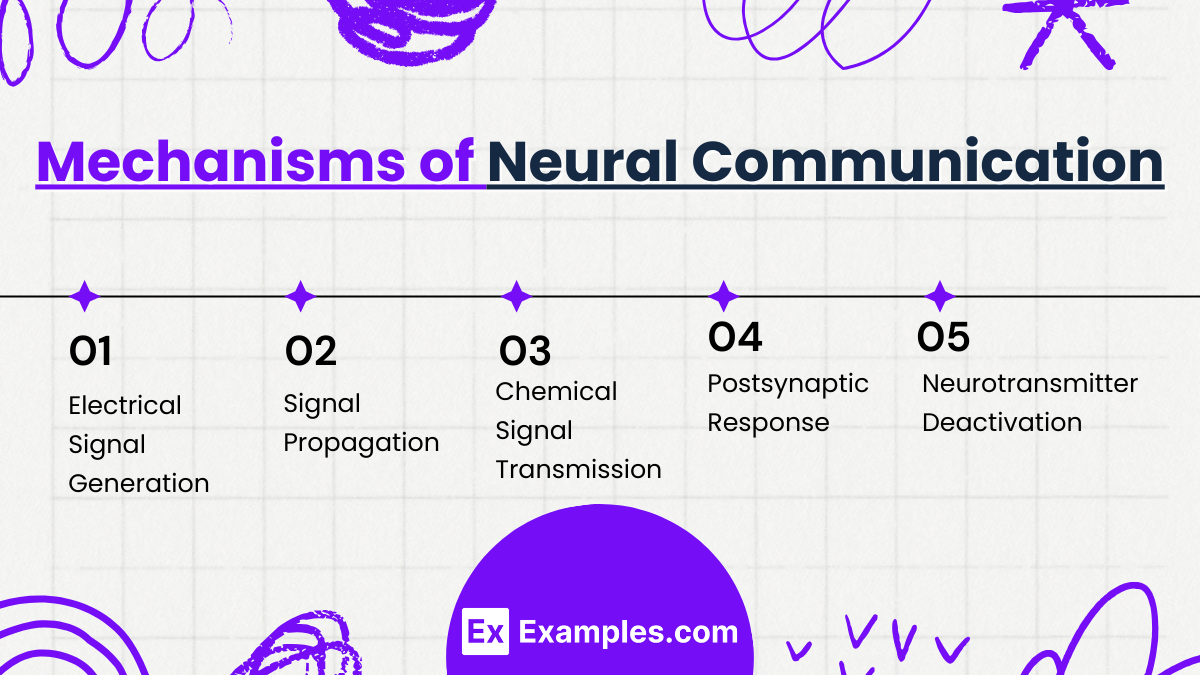
Neural communication is a complex process that allows neurons to transmit information throughout the body. Here's a simplified overview of the mechanisms involved:
1. Electrical Signal Generation
Resting Potential: Neurons maintain a resting membrane potential due to the differential distribution of ions across their membranes, typically around -70 mV.
Action Potential: When a neuron is stimulated by signals, if the stimulation reaches a certain threshold, it triggers an action potential. This is a rapid, temporary change in the membrane potential, where the inside of the neuron becomes positively charged compared to the outside.
2. Signal Propagation
Axon Conduction: The action potential travels along the axon from the cell body to the axon terminals. This propagation is facilitated by the myelin sheath in many neurons, which increases the speed of transmission through a process called saltatory conduction.
3. Chemical Signal Transmission
Synaptic Transmission: At the end of the axon, the action potential reaches the synapse, a small gap between neurons.
Neurotransmitter Release: The arrival of an action potential triggers the opening of voltage-gated calcium channels, causing neurotransmitters (chemical messengers) stored in vesicles to be released into the synaptic cleft.
Receptor Binding: These neurotransmitters cross the synaptic cleft and bind to specific receptors on the postsynaptic neuron’s membrane, initiating a response.
4. Postsynaptic Response
Excitatory or Inhibitory: Depending on the type of neurotransmitter and receptor involved, the binding may cause an excitatory or inhibitory postsynaptic potential. Excitatory inputs increase the likelihood of firing an action potential, while inhibitory inputs decrease it.
Signal Integration: The postsynaptic neuron integrates all excitatory and inhibitory signals. If the net effect is excitatory and surpasses the threshold, it will fire an action potential, continuing the communication process.
5. Neurotransmitter Deactivation
Reuptake, Enzymatic Degradation, or Diffusion: After the neurotransmitter has acted, it is either taken back into the presynaptic neuron, degraded by enzymes, or diffused out of the synaptic cleft, terminating the signal.
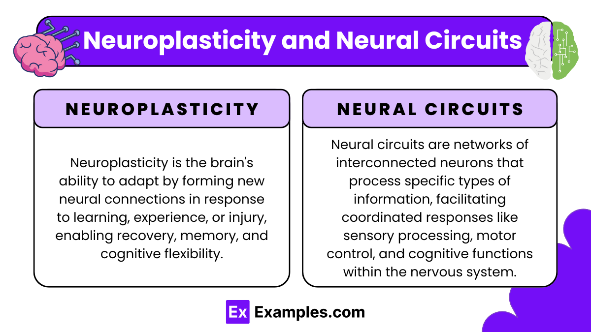
Neuroplasticity and Neural Circuits
Neuroplasticity and neural circuits are fundamental concepts in understanding how the brain adapts and functions. Here’s a simplified explanation of both:
Neuroplasticity
Neuroplasticity refers to the brain's ability to reorganize itself by forming new neural connections throughout life. This adaptability is a response to learning, experience, or injury. Key aspects include:
Synaptic Plasticity: The strength of synaptic connections between neurons can increase (potentiation) or decrease (depression) over time, reflecting changes in behavior and learning.
Structural Plasticity: This involves changes in the physical structure of the brain, such as new synapses forming or the pruning of neurons that are no longer needed.
Functional Plasticity: After damage, the brain can transfer functions from a damaged area to undamaged areas, demonstrating its ability to reassign tasks.
Neural Circuits
Neural circuits are networks of neurons connected by synapses that work together to carry out a specific function or behavior. These circuits can be simple, like those governing reflexes, or complex, involving vast areas of the brain for higher-order functions. Key points include:
Circuit Formation: Neural circuits are formed through the process of synaptic connections between neurons, guided by both genetic factors and experience.
Circuit Modulation: Existing circuits can be modified by incoming sensory experiences or by other circuits, which can inhibit or stimulate their function.
Feedback Loops: Many neural circuits include feedback mechanisms that help regulate and refine their output, important for maintaining balance and appropriate responses.
Neural Cell Dysfunction and Neurological Disorders
Neural cell dysfunction and its association with neurological disorders encompass a broad range of conditions that affect the brain, spinal cord, and nerves. Here’s an overview of how neural cell dysfunction leads to neurological disorders and some common examples:
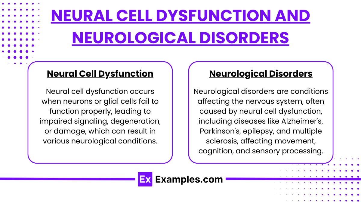
Neural Cell Dysfunction
Neural cell dysfunction occurs when neurons or glial cells fail to function properly, leading to impaired signaling, degeneration, or damage, which can result in various neurological conditions.. This can be due to genetic defects, environmental factors, trauma, or disease, leading to various neurological disorders. Key types of dysfunction include:
Loss of Neurotransmission: Impaired synaptic function can lead to reduced signaling efficiency between neurons, affecting brain function and leading to diseases such as Parkinson’s disease, where dopamine production is diminished.
Neurodegeneration: The progressive loss of neuron structure or function, including death of neurons, is characteristic of diseases like Alzheimer’s and Huntington’s disease.
Demyelination: The loss of the myelin sheath surrounding axons disrupts signal transmission, as seen in multiple sclerosis.
Neuroinflammation: Chronic inflammation within neural tissues can lead to or exacerbate disorders like epilepsy and Alzheimer’s disease.
Excitotoxicity: Excessive neurotransmitter release or ineffective reuptake can lead to overactivation of receptors, particularly glutamate receptors, which can cause neuron damage and death.
Neurological Disorders
Neurological disorders are conditions affecting the nervous system, often caused by neural cell dysfunction, including diseases like Alzheimer's, Parkinson's, epilepsy, and multiple sclerosis, affecting movement, cognition, and sensory processing.
Here are some disorders associated with neural cell dysfunction:
Alzheimer’s Disease: Characterized by neurodegeneration and the accumulation of amyloid-beta plaques and tau tangles, leading to memory loss and cognitive decline.
Parkinson’s Disease: A movement disorder resulting from the death of dopamine-producing neurons in the brain, leading to tremors, rigidity, and bradykinesia (slowed movement).
Multiple Sclerosis (MS): An autoimmune disorder where the immune system attacks myelin in the CNS, leading to impaired motor function, sensory symptoms, and cognitive challenges.
Amyotrophic Lateral Sclerosis (ALS): Also known as Lou Gehrig’s disease, ALS involves the degeneration of motor neurons, leading to muscle weakness and eventual loss of voluntary muscle control.
Epilepsy: Caused by abnormal electrical brain activity, leading to seizures. It can result from genetic conditions, brain injury, or unknown factors.
Examples
Example 1:Neuron Action Potential
A sensory neuron in the skin detects a painful stimulus.
The neuron's membrane potential changes, triggering an action potential that rapidly travels along the neuron's axon to the spinal cord, where it initiates a reflex response while also conveying the pain signal to the brain.
Example 2:Synaptic Plasticity in Learning
Learning a new motor skill, like playing the piano.
Repeated practice strengthens synaptic connections in the motor cortex associated with finger movements. This synaptic strengthening, known as long-term potentiation (LTP), enhances the efficiency of neuron firing, making the skill easier and more automatic over time.
Example 3:Glial Cell Function in the CNS
Following a neuronal injury.
Microglia (a type of glial cell) activate to clean up debris and dead cells at the injury site, while astrocytes contribute to the formation of a scar and help restore the blood-brain barrier, protecting remaining healthy neural tissue.
Example 4:Neurotransmitter Dysfunction in Depression
An imbalance in serotonin levels in the brain.
Lower than normal levels of serotonin in synaptic clefts can lead to symptoms of depression. Antidepressants like SSRIs (Selective Serotonin Reuptake Inhibitors) work by blocking the reuptake of serotonin, increasing its availability in the brain, thereby improving mood and emotion regulation.
Example 5:Neurodegenerative Disease Progression
Parkinson’s disease affecting motor control.
Loss of dopaminergic neurons in the substantia nigra part of the brain leads to reduced dopamine levels, critical for regulating movement. This reduction causes symptoms like tremors, rigidity, and bradykinesia. Treatment often includes medications that increase dopamine levels or mimic dopamine effects.
Practice Questions
Question 1:
What is primarily responsible for the resting potential of a neuron?
A) The high concentration of sodium ions inside the cell
B) The high concentration of potassium ions inside the cell
C) The selective permeability of the neuronal membrane to potassium ions
D) The action of sodium-potassium pumps in expelling potassium from the cell
Answer: C) The selective permeability of the neuronal membrane to potassium ions
Explanation:
The resting potential of a neuron is largely due to the selective permeability of its membrane to potassium ions. While sodium-potassium pumps do play a role in maintaining ion concentration gradients, it is the potassium channels that allow potassium to flow out of the cell more than sodium flows in, thus establishing a negative internal environment typical of a resting neuron.
Question 2:
Which neurotransmitter is primarily involved in the excitation of muscles and is also important for memory and learning in the brain?
A) Dopamine
B) Serotonin
C) Acetylcholine
D) GABA
Answer: C) Acetylcholine
Explanation:
Acetylcholine is crucial for muscle activation at neuromuscular junctions where it stimulates muscle contractions. In the brain, acetylcholine is involved in arousal, attention, memory, and motivation, making it essential for both peripheral and central nervous system functions.
Question 3:
Which glial cell type is responsible for myelinating neurons in the central nervous system?
A) Astrocytes
B) Oligodendrocytes
C) Schwann cells
D) Microglia
Answer: B) Oligodendrocytes
Explanation:
Oligodendrocytes are the glial cells responsible for myelinating neurons in the central nervous system. Each oligodendrocyte can extend its processes to several axons, wrapping them with a myelin sheath that speeds up the electrical transmission along the neuron. In contrast, Schwann cells provide myelination in the peripheral nervous system.