
Neuronal Synapses

Preparing for the MCAT requires a solid understanding of neuronal synapses, as they are fundamental to neurology within organ systems. Synapses facilitate neuron-to-neuron communication through neurotransmitters, vital for transmitting signals that influence everything from muscle contractions to cognition. Understanding these interactions is crucial for mastering the nervous system's role in health and disease.
Learning Objective
In studying "Neuronal Synapses" for the MCAT, you should aim to grasp the complex mechanisms of synaptic transmission and neuronal communication. Examine the structural components of synapses, including presynaptic terminals, synaptic clefts, and postsynaptic receptors. Understand the role of neurotransmitters and their receptors in signal propagation and modulation. Explore synaptic plasticity, including long-term potentiation and depression, which underpin learning and memory. Evaluate how disruptions in synaptic function can lead to neurological disorders. Apply this knowledge to analyze and interpret neurophysiological data and scenarios presented in MCAT practice passages.
Introduction to Neuronal Synapses
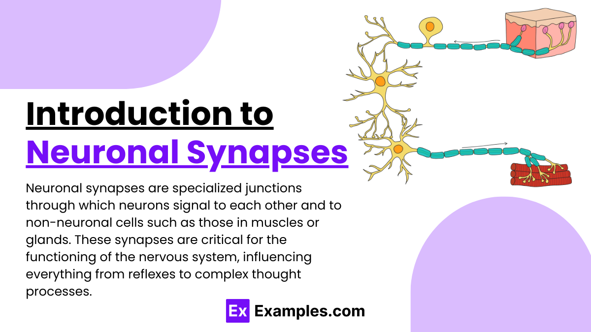
Neuronal synapses are specialized junctions through which neurons signal to each other and to non-neuronal cells such as those in muscles or glands. These synapses are critical for the functioning of the nervous system, influencing everything from reflexes to complex thought processes. Here’s a basic overview of how they work:
Types of Synapses
Electrical Synapses: These synapses directly pass ions through gap junctions, allowing rapid synchronization of electrical activity among neurons. They are less common than chemical synapses.
Chemical Synapses: These are more common and involve the release of neurotransmitters from the presynaptic neuron. When an action potential reaches the synaptic terminal, it triggers the release of these chemicals, which cross the synaptic cleft and bind to receptors on the postsynaptic neuron, initiating a new electrical signal.
Key Components of a Synapse
Presynaptic Terminal: The end of the neuron that releases neurotransmitters.
Synaptic Cleft: A small gap between the presynaptic and postsynaptic neurons where neurotransmitters are released.
Postsynaptic Terminal: The part of the neuron that receives the neurotransmitter and generates a response, typically through receptor molecules.
Mechanism of Synaptic Transmission
Action Potential Arrival: An action potential arrives at the presynaptic terminal.
Neurotransmitter Release: The action potential causes calcium ions to enter the presynaptic neuron, triggering the release of neurotransmitters stored in vesicles.
Neurotransmitter Binding: The neurotransmitters cross the synaptic cleft and bind to specific receptors on the postsynaptic neuron.
Postsynaptic Potential: This binding opens ion channels, leading to changes in the postsynaptic neuron’s membrane potential.
Signal Propagation or Inhibition: Depending on the type of neurotransmitter and receptor involved, the postsynaptic neuron may be excited or inhibited, leading to either the continuation or suppression of the action potential.
Structural Components of Synapses
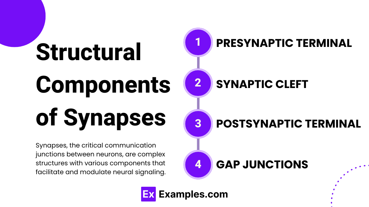
Synapses, the critical communication junctions between neurons, are complex structures with various components that facilitate and modulate neural signaling. Here’s a detailed breakdown of the structural components that constitute both chemical and electrical synapses:
Components of Chemical Synapses
Presynaptic Terminal (Axon Terminal):
Synaptic Vesicles: Small membrane-bound structures that store neurotransmitters, the chemical messengers of the nervous system.
Voltage-Gated Calcium Channels: These channels open in response to action potentials, allowing calcium ions to enter the neuron, which is crucial for neurotransmitter release.
Active Zone: A specialized area within the presynaptic terminal where neurotransmitters are released. It contains proteins that regulate neurotransmitter release.
Synaptic Cleft:
This is the narrow gap between the presynaptic and postsynaptic neurons, typically about 20-40 nanometers wide. The synaptic cleft is filled with extracellular matrix proteins that help stabilize the synaptic connection and guide the diffusion of neurotransmitters.
Postsynaptic Terminal:
Neurotransmitter Receptors: Proteins on the postsynaptic neuron’s membrane that bind neurotransmitters and initiate a response in the neuron.
Postsynaptic Density: A dense network of proteins located directly opposite the active zone in the presynaptic neuron. It contains receptors, structural proteins, and signaling enzymes that transduce and regulate synaptic signals.
Ion Channels: Proteins that allow ions to enter or exit the neuron, changing the neuron's electrical charge and triggering a response.
Components of Electrical Synapses
Gap Junctions:
Connexons: These are channels that connect adjacent neurons directly at electrical synapses. Each connexon is formed from six connexin proteins in one neuron aligning with six connexins from an adjacent neuron to form a conduit for ions and small molecules.
Unlike chemical synapses, electrical synapses allow for the direct passage of ionic currents from one neuron to another, enabling faster communication and synchronization.
Neurotransmitters and Signal Transmission
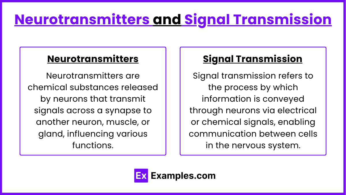
Neurotransmitters
Neurotransmitters are chemical messengers that facilitate communication between neurons and other cells. They vary widely in type, including small molecules like dopamine and serotonin, peptides such as endorphins, and gases like nitric oxide. Produced and stored in neurons, neurotransmitters are released in response to electrical signals, cross the synaptic gap, and bind to receptors on the postsynaptic cell, triggering further cellular responses. Their action concludes with their reuptake, degradation, or absorption by nearby cells to terminate the signal.
Signal Transmission
Signal transmission at synapses begins when an action potential reaches a neuron's axon terminal, prompting the opening of voltage-gated calcium channels. The influx of calcium causes neurotransmitters to be released into the synaptic cleft. These neurotransmitters then bind to receptors on the postsynaptic neuron, causing ion channels to open and modify the neuron's membrane potential, which can excite or inhibit the neuron depending on the type of neurotransmitter and receptor involved. The process ends when neurotransmitters are cleared from the synaptic cleft through reuptake by the presynaptic neuron, degradation by enzymes, or absorption by glial cells. This cycle is essential for neural communication and overall nervous system function.
Synaptic plasticity is the ability of synapses to strengthen or weaken over time in response to increases or decreases in their activity. This dynamic process is essential for learning, memory, and the adaptation of the brain to new information. Here’s a streamlined explanation of how synaptic plasticity works:
Synaptic Plasticity

Synaptic plasticity is the ability of synapses, the connections between neurons, to strengthen or weaken over time in response to increases or decreases in activity. This adaptability is crucial for learning, memory formation, and the brain’s ability to reorganize following injury or experience.
Mechanisms of Synaptic Plasticity
Long-Term Potentiation (LTP): LTP is one of the most well-studied forms of synaptic plasticity, characterized by a long-lasting increase in synaptic strength following a high-frequency stimulation of the synapse. This process involves the enhanced release of neurotransmitters, increased responsiveness of postsynaptic neurons to neurotransmitters, and changes in receptor density and function.
Long-Term Depression (LTD): In contrast to LTP, LTD involves a long-lasting decrease in synaptic strength, typically following low-frequency stimulation of the synapse. This reduction in synaptic strength is crucial for deleting old memories and can prevent the overloading of neural circuits.
Molecular Basis
NMDA Receptor Activation: Both LTP and LTD can be dependent on the activation of NMDA (N-methyl-D-aspartate) receptors, which are sensitive to the intensity and timing of synaptic activity. Calcium influx through NMDA receptors plays a pivotal role, triggering intracellular signaling pathways that modify synaptic strength.
Changes at the Synapse: Depending on the signals received, the synapse may undergo structural changes such as the alteration of the number and size of dendritic spines (small protrusions on dendrites where synapses are located), and the arrangement of molecules within the synapse that affect how effectively neurons communicate.
Examples
Example 1:Synaptic Transmission
A signal needs to pass from one neuron to another in the central nervous system.
This occurs when an action potential reaches the presynaptic terminal, causing calcium ions to enter the cell, which triggers the release of neurotransmitters into the synaptic cleft. These neurotransmitters bind to receptors on the postsynaptic neuron, causing a new action potential to begin if the signal is strong enough.
Example 2:Neurotransmitter Reuptake
Serotonin transmission in the synaptic cleft.
After serotonin is released into the synaptic cleft and binds to its receptors, it is taken back into the presynaptic neuron by serotonin transporters. This process is targeted by many antidepressant drugs to increase serotonin levels in the brain, affecting mood and anxiety levels.
Example 3:Long-term Potentiation (LTP)
Enhancing synaptic connections during learning.
LTP occurs when repeated stimulation of a synapse results in a sustained increase in the amplitude of postsynaptic potentials. This enhancement can last from hours to weeks and is a cellular basis for learning and memory.
Example 4:Synaptic Plasticity Disruption in Disease
Alzheimer’s disease impacts synaptic connections.
In Alzheimer's, abnormal levels of amyloid beta peptide lead to synaptic loss and dysfunction. This disrupts neural communication and results in memory loss, cognitive decline, and changes in behavior.
Example 5:Inhibition of Neurotransmitter Release
The effect of botulinum toxin on neuromuscular junctions.
Botulinum toxin prevents the release of acetylcholine, a neurotransmitter at neuromuscular junctions, effectively causing muscle paralysis. This mechanism is exploited clinically for treatments of muscle spasms and cosmetically to reduce wrinkles by temporarily paralyzing facial muscles.
Practice Questions
Question 1:
What is the primary function of neurotransmitters at a chemical synapse?
A) To stimulate the release of calcium ions in the presynaptic neuron
B) To modify the DNA transcription in the postsynaptic neuron
C) To transmit signals between neurons by binding to receptors
D) To facilitate the passive transport of ions across the synaptic cleft
Answer: C) To transmit signals between neurons by binding to receptors
Explanation: Neurotransmitters are chemicals released from the synaptic vesicles in the presynaptic neuron into the synaptic cleft. Their primary function is to transmit signals between neurons by binding to specific receptors on the postsynaptic neuron, which can then initiate a new action potential or modulate neuronal activity, depending on the type of neurotransmitter and receptor involved.
Question 2:
Which of the following is true about synaptic plasticity?
A) It only occurs in the motor areas of the brain
B) It is the process by which synapses decrease their effectiveness in response to frequent use
C) It involves changes in synaptic strength in response to levels of activity
D) It permanently eliminates unnecessary synapses at birth
Answer: C) It involves changes in synaptic strength in response to levels of activity
Explanation: Synaptic plasticity refers to the ability of synapses to strengthen or weaken over time in response to increases or decreases in their activity. This dynamic change in synaptic strength is crucial for learning, memory, and adaptation to new information or environments, and it does not exclusively occur in any single brain area nor does it involve the permanent elimination of synapses at birth.
Question 3:
What is the role of calcium ions in synaptic transmission?
A) They deactivate neurotransmitter molecules in the synaptic cleft
B) They trigger the fusion of vesicles with the presynaptic membrane to release neurotransmitters
C) They are released into the synaptic cleft to directly stimulate the postsynaptic neuron
D) They prevent the reuptake of neurotransmitters by the presynaptic neuron
Answer: B) They trigger the fusion of vesicles with the presynaptic membrane to release neurotransmitters
Explanation: Calcium ions play a crucial role in synaptic transmission by entering the presynaptic neuron through voltage-gated calcium channels upon the arrival of an action potential. The influx of calcium ions triggers a cascade of events that lead to the fusion of neurotransmitter-containing synaptic vesicles with the presynaptic membrane. This fusion releases neurotransmitters into the synaptic cleft, enabling them to bind to receptors on the postsynaptic neuron and propagate the neural signal.