
Prokaryotes/Bacteria

Preparing for the MCAT requires a thorough understanding of prokaryotes, particularly bacteria, a key component of the Biological and Biochemical Foundations section. Mastery of bacterial structure, growth, and genetic mechanisms is essential. This knowledge provides insights into microbial behavior, antibiotic resistance, and human health, critical for achieving a high MCAT score.
Learning Objective
In studying "Prokaryotes/Bacteria" for the MCAT, you should understand the structural and functional differences between prokaryotic and eukaryotic cells. Analyze bacterial cell structures such as cell walls, capsules, flagella, and pili, and their roles in survival, motility, and pathogenicity. Evaluate bacterial growth, replication (binary fission), and genetic exchange mechanisms like conjugation, transformation, and transduction. Additionally, explore the role of bacteria in human health, including beneficial gut flora and pathogenic bacteria, and the principles behind antibiotic mechanisms and resistance. Apply this understanding to interpret experimental data and scenarios in MCAT practice passages, enhancing your preparedness for exam questions.
Overview of Prokaryotic Cell Structure
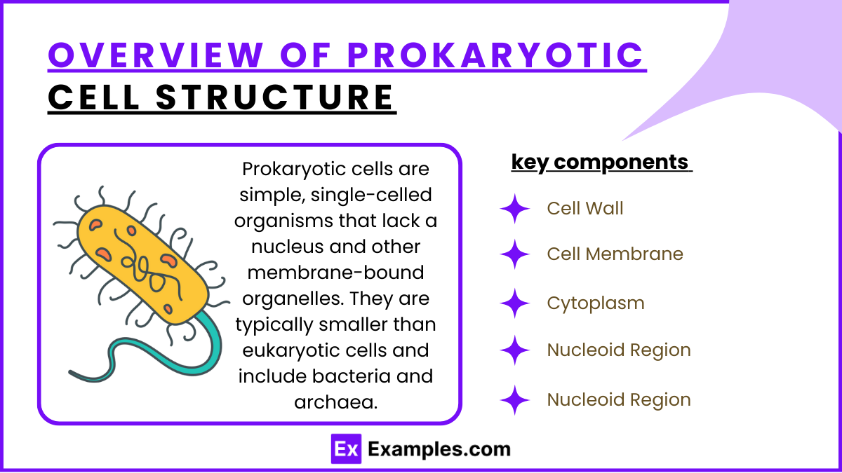
Prokaryotic cells are simple, single-celled organisms that lack a nucleus and other membrane-bound organelles. They are typically smaller than eukaryotic cells and include bacteria and archaea. Below are the key components of a prokaryotic cell:
1. Cell Wall
Provides structural support and protection.
Composed of peptidoglycan (in bacteria) or other polymers (in archaea).
Maintains cell shape and prevents it from bursting in hypotonic environments.
2. Cell Membrane
A lipid bilayer that controls the movement of substances in and out of the cell.
Contains proteins involved in energy production, nutrient transport, and cell communication.
3. Cytoplasm
Gel-like substance where cellular components and enzymes are located.
Contains ribosomes, which are involved in protein synthesis.
4. Nucleoid Region
The area where the cell’s genetic material (a single circular DNA chromosome) is located.
Not enclosed by a membrane, unlike the nucleus in eukaryotic cells.
5. Ribosomes
Smaller than eukaryotic ribosomes (70S vs. 80S).
Responsible for synthesizing proteins based on the genetic information in the DNA.
Bacterial Growth and Replication
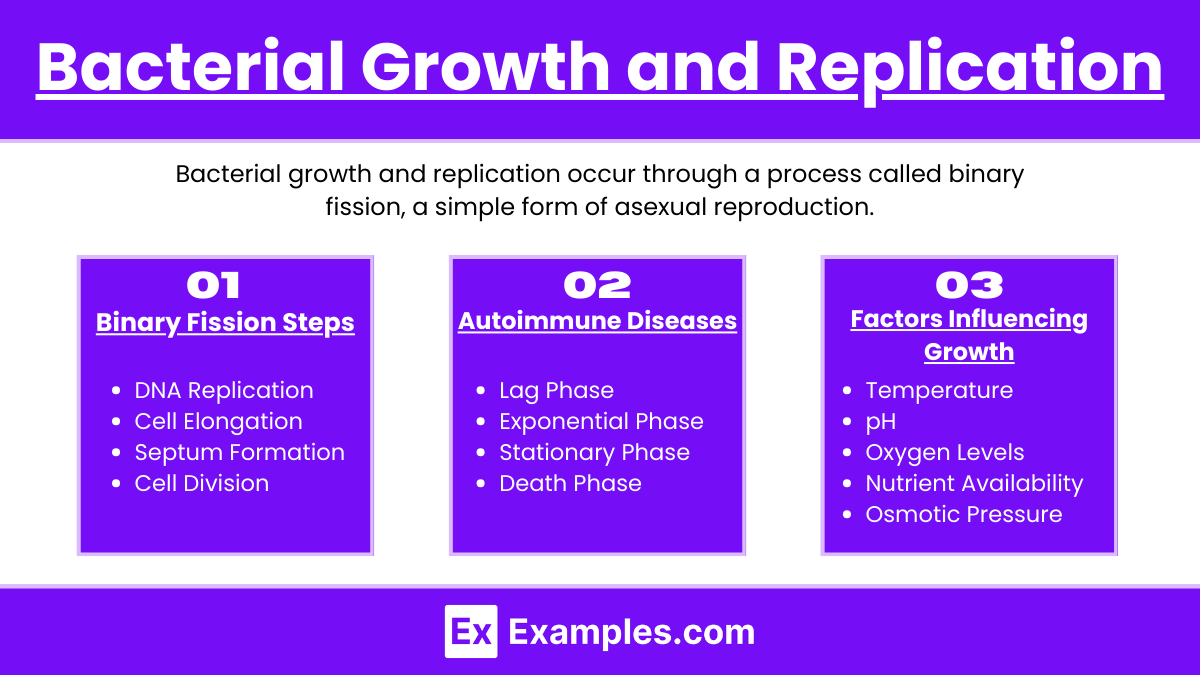
Bacterial growth and replication occur through a process called binary fission, a simple form of asexual reproduction. This process and the conditions that affect bacterial growth are outlined below:
1. Binary Fission Steps:
DNA Replication: The single bacterial chromosome is copied.
Cell Elongation: The cell grows, separating the DNA copies.
Septum Formation: A wall forms in the middle of the cell.
Cell Division: The cell splits into two identical cells.
2. Phases of Bacterial Growth:
Lag Phase: Bacteria adapt; no increase in number.
Exponential Phase: Rapid cell division and growth.
Stationary Phase: Growth slows as resources run low.
Death Phase: Cells die due to nutrient depletion and waste accumulation.
3. Factors Influencing Growth:
Temperature: Each bacterial type has an optimal range.
pH: Most bacteria prefer neutral conditions.
Oxygen Levels:
Aerobes need oxygen.
Anaerobes grow without oxygen.
Facultative Anaerobes can grow with or without oxygen.
Nutrient Availability: Essential for growth.
Osmotic Pressure: Balanced conditions are necessary.
Genetic Exchange Mechanisms
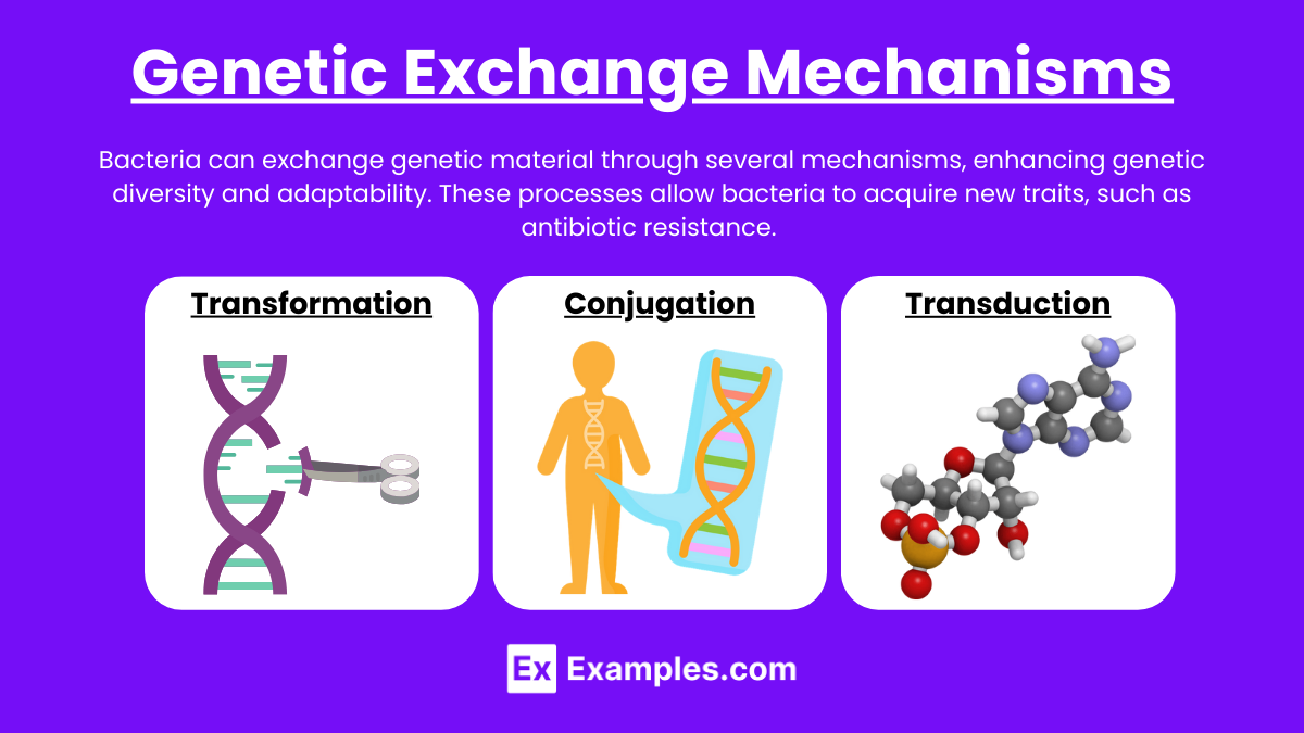
Bacteria can exchange genetic material through several mechanisms, enhancing genetic diversity and adaptability. These processes allow bacteria to acquire new traits, such as antibiotic resistance. The main genetic exchange mechanisms are:
1. Transformation
Bacteria take up free DNA fragments from their environment, often from dead bacteria.
The foreign DNA can integrate into the bacterial genome, giving the cell new characteristics.
2. Conjugation
Involves direct cell-to-cell contact through a structure called a pilus.
A donor bacterium transfers a plasmid (a small, circular DNA molecule) to a recipient cell.
The plasmid often carries genes that provide advantages, such as antibiotic resistance.
3. Transduction
A bacteriophage (a virus that infects bacteria) carries DNA from one bacterium to another.
During infection, the phage may accidentally package bacterial DNA and transfer it when infecting a new host.
This transferred DNA can recombine with the recipient bacterium’s genome.
Experimental Techniques
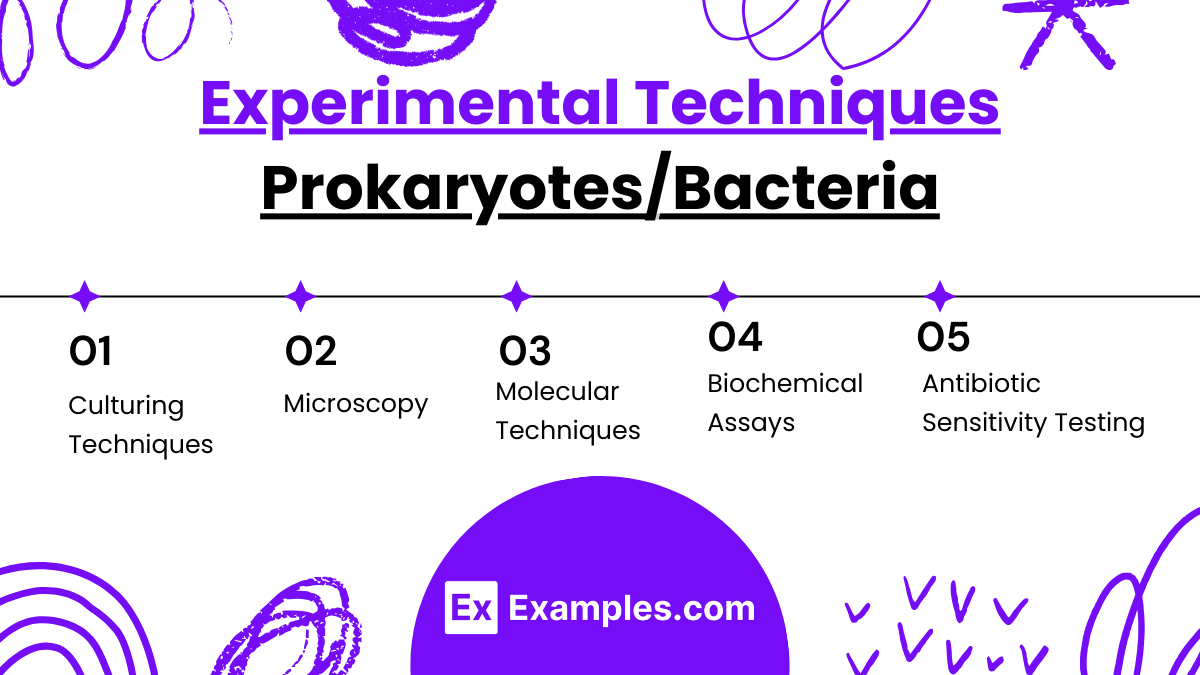
Studying prokaryotes, particularly bacteria, involves various experimental techniques that are crucial for research, medicine, and biotechnology. These methods allow scientists to understand bacterial functions, identify pathogens, and develop new applications.
1. Culturing Techniques
Aseptic Technique: Ensures bacteria are cultured in a sterile environment to prevent contamination.
Growth Media: Bacteria are grown on agar plates or in liquid media to study their growth, morphology, and colony characteristics.
Selective and Differential Media: Used to isolate specific bacterial strains or distinguish between different types based on biochemical properties.
2. Microscopy
Light Microscopy: Visualizes bacteria to examine shape, size, and motility.
Fluorescence Microscopy: Stains bacteria with fluorescent dyes or labels specific proteins, enabling the observation of cellular structures and interactions.
Electron Microscopy: Provides high-resolution images of bacterial ultrastructure, revealing detailed cellular components.
3. Molecular Techniques
PCR (Polymerase Chain Reaction): Amplifies bacterial DNA for identification, genetic studies, or detecting pathogens.
DNA Sequencing: Determines the genetic sequence of bacteria, helping identify species and genetic variations (e.g., antibiotic resistance genes).
CRISPR-Cas9: A gene-editing tool used to manipulate bacterial genes for research or biotechnological applications.
4. Biochemical Assays
ELISA (Enzyme-Linked Immunosorbent Assay): Detects specific bacterial antigens or antibodies in samples, often used for diagnosing infections.
Metabolic Tests: Assesses bacterial metabolic capabilities (e.g., carbohydrate fermentation tests) to identify species or study biochemical pathways.
5. Antibiotic Sensitivity Testing
Determines the effectiveness of antibiotics against bacterial strains.
Methods like the Kirby-Bauer Disk Diffusion Test evaluate the inhibition zones around antibiotic disks, indicating bacterial susceptibility or resistance.
Examples
Examples 1: Gram-Positive vs. Gram-Negative Bacteria
Gram-positive bacteria have a thick peptidoglycan layer that retains the crystal violet stain, while gram-negative bacteria have a thin peptidoglycan layer and an outer membrane, making them more resistant to certain antibiotics. This distinction is important in clinical diagnosis and treatment.
Examples 2: Binary Fission as a Mode of Replication
Escherichia coli (E. coli) rapidly divides by binary fission, doubling in number every 20 minutes under optimal conditions. This illustrates how bacterial populations can grow exponentially, impacting infection spread and antibiotic treatment strategies.
Examples 3: Antibiotic Resistance through Conjugation
Bacteria like Staphylococcus aureus can acquire antibiotic resistance genes through conjugation, where a plasmid carrying resistance genes is transferred from one bacterium to another. Understanding this mechanism is crucial for developing strategies to combat antibiotic resistance.
Examples 4: Endospore Formation in Bacillus and Clostridium
Bacillus and Clostridium species form endospores to survive harsh conditions, such as extreme heat or lack of nutrients. These endospores can remain dormant for long periods and are resistant to disinfection, posing challenges in sterilization and infection control.
Examples 5: The Role of the Human Gut Microbiome
Beneficial bacteria such as Lactobacillus and Bifidobacterium play a key role in digestion, vitamin synthesis, and immune function. Disruptions in the gut microbiome, like those caused by antibiotics, can lead to digestive issues and immune imbalances, highlighting the importance of maintaining a healthy microbial balance.
Practice Questions:
Question 1
Which of the following structures is found in prokaryotic cells but not in eukaryotic cells?
A) Nucleus
B) Mitochondria
C) Peptidoglycan cell wall
D) Endoplasmic reticulum
Answer: C) Peptidoglycan cell wall
Explanation:
Prokaryotic cells, specifically bacteria, have a cell wall composed of peptidoglycan, which provides structural support and protection. Eukaryotic cells do not have peptidoglycan in their cell walls (if they have cell walls at all, such as in plants). Eukaryotic cells possess organelles like the nucleus, mitochondria, and endoplasmic reticulum, which prokaryotic cells lack.
Question 2
Which of the following processes is primarily responsible for genetic exchange between bacteria?
A) Binary fission
B) Mitosis
C) Conjugation
D) Meiosis
Answer: C) Conjugation
Explanation:
Conjugation is a process where bacteria transfer genetic material through a pilus, enabling the spread of traits such as antibiotic resistance. Binary fission is how bacteria reproduce asexually, while mitosis and meiosis are processes associated with eukaryotic cell division and are not found in prokaryotes.
Question 3
Which type of bacteria is characterized by a thin layer of peptidoglycan and an outer membrane, making it resistant to certain antibiotics?
A) Gram-positive bacteria
B) Gram-negative bacteria
C) Acid-fast bacteria
D) Spirochetes
Answer: B) Gram-negative bacteria
Explanation:
Gram-negative bacteria have a thin layer of peptidoglycan located between an inner and outer membrane. The outer membrane contains lipopolysaccharides and acts as a barrier to certain antibiotics, making these bacteria generally more resistant. Gram-positive bacteria, in contrast, have a thick peptidoglycan layer but lack an outer membrane, while acid-fast bacteria and spirochetes have different structural characteristics.