Preparing for the MCAT requires a solid understanding of protein analysis techniques, as they play a crucial role in molecular biology and biochemistry. Key techniques include chromatography, mass spectrometry, Western blotting, and ELISA, each providing insights into protein structure, function, and interactions. Mastery of these methods will enhance your grasp of complex biological systems, aiding in achieving a high MCAT score.
Learning Objective
In studying “Protein Analysis Techniques” for the MCAT, you should learn to understand the various methods used to analyze proteins, including electrophoresis, chromatography, and mass spectrometry. Analyze how these techniques help determine protein size, structure, and function. Evaluate the principles behind techniques like Western blotting, enzyme-linked immunosorbent assay (ELISA), and X-ray crystallography. Additionally, explore how these tools are used in both research and clinical applications to study protein interactions, modifications, and activity, and apply your understanding to interpreting experimental results in MCAT practice passages.
1. Protein Purification

Protein purification involves isolating a specific protein from a complex mixture for study or use. Techniques like chromatography, centrifugation, and electrophoresis are commonly employed to achieve purity.
- Techniques:
- Affinity Chromatography: Uses specific ligands to bind the target protein, separating it based on unique binding interactions.
- Ion Exchange Chromatography: Separates proteins based on charge; cation and anion exchange resins attract proteins with opposite charges.
- Gel Filtration (Size Exclusion) Chromatography: Separates proteins by size, with larger proteins eluting first as they cannot enter the pores of the matrix.
2. Electrophoresis
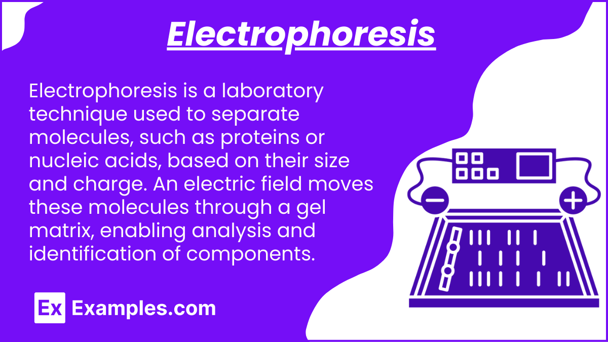
Electrophoresis is a laboratory technique used to separate molecules, such as proteins or nucleic acids, based on their size and charge. An electric field moves these molecules through a gel matrix, enabling analysis and identification of components.
- Techniques:
- SDS-PAGE (Sodium Dodecyl Sulfate-Polyacrylamide Gel Electrophoresis): Denatures proteins, giving them a uniform negative charge and allowing separation based solely on size.
- Native PAGE: Maintains proteins’ native structure, allowing separation based on charge, size, and shape.
- Isoelectric Focusing (IEF): Separates proteins by isoelectric point (pI) in a pH gradient, where proteins stop migrating when pH = pI.
- 2D Gel Electrophoresis: Combines IEF and SDS-PAGE for separation based on both pI and molecular weight, offering high-resolution separation.
3. Enzyme-Linked Immunosorbent Assay (ELISA)

ELISA is a widely used technique for detecting and quantifying specific proteins or antigens in a sample, leveraging the high specificity of antibodies. It is used in diagnostics, biomedical research, and various biological assays.
- Sandwich ELISA:
- This method is typically used when the target protein is a large molecule with multiple binding sites. It uses two antibodies: a capture antibody bound to the plate and a detection antibody that binds to a different site on the protein.
- Process: The sample is added to a plate with the capture antibody, which binds to the target protein. After washing to remove non-specific binding, a detection antibody conjugated to an enzyme is added. The enzyme’s activity produces a detectable signal, proportional to the protein amount.
- Competitive ELISA:
- Useful for smaller proteins or when only one binding site is available. In this format, the sample competes with a labeled version of the protein for binding to the capture antibody.
- Process: The more protein present in the sample, the less labeled protein binds to the antibody. The signal is inversely proportional to the protein concentration.
4. Mass Spectrometry (MS)
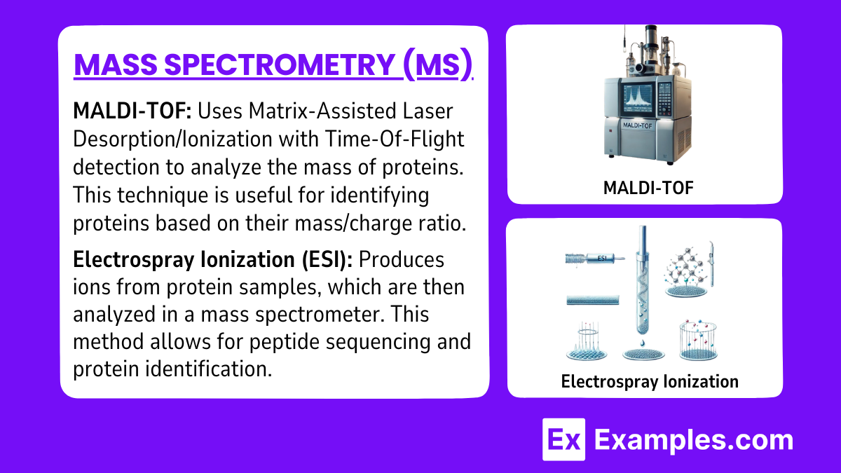
- MALDI-TOF: Uses Matrix-Assisted Laser Desorption/Ionization with Time-Of-Flight detection to analyze the mass of proteins. This technique is useful for identifying proteins based on their mass/charge ratio.
- MALDI-TOF is a mass spectrometry technique that combines Matrix-Assisted Laser Desorption/Ionization (MALDI) with Time-Of-Flight (TOF) detection. It is particularly useful for analyzing biomolecules like proteins and peptides, which are typically challenging to ionize and analyze by traditional means.
- Electrospray Ionization (ESI): Produces ions from protein samples, which are then analyzed in a mass spectrometer. This method allows for peptide sequencing and protein identification.
- ESI is a soft ionization technique, ideal for analyzing large biomolecules like proteins and peptides. It is often used in conjunction with tandem mass spectrometry (MS/MS) to gain detailed structural information, including peptide sequencing and the identification of post-translational modifications.
Examples
Example 1: Western Blotting
This technique involves separating proteins based on size using SDS-PAGE, followed by transferring them onto a membrane. Specific proteins are then detected using antibodies that bind to the target protein. Western blotting is widely used to confirm the presence of a protein and assess its relative abundance. It’s commonly applied in research areas like molecular biology and diagnostics for diseases such as cancer and viral infections.
Example 2: ELISA (Enzyme-Linked Immunosorbent Assay)
ELISA is used to detect and quantify specific proteins or antigens in a sample. This method involves an antibody that binds to the target protein and an enzyme that produces a colorimetric or fluorescent signal. Variants such as sandwich and competitive ELISA allow flexibility depending on the protein’s properties and concentration. ELISA is commonly used in immunology, disease diagnostics, and vaccine development.
Example 3: Mass Spectrometry (MS)
Mass spectrometry is a highly sensitive technique for identifying proteins and analyzing their structure. Techniques like MALDI-TOF and ESI are commonly used to ionize proteins and measure their mass-to-charge ratios, allowing researchers to determine the composition and sequence of proteins. Mass spectrometry is crucial for proteomics, drug development, and biomarker discovery due to its precision and versatility.
Example 4: X-ray Crystallography
This method is used to determine the three-dimensional structure of proteins at the atomic level. Crystallized proteins are bombarded with X-rays, producing diffraction patterns that reveal the spatial arrangement of atoms. X-ray crystallography is essential for understanding protein functions, enzyme mechanisms, and aiding in drug design by providing detailed structural insights.
Example 5: NMR (Nuclear Magnetic Resonance) Spectroscopy
NMR spectroscopy is ideal for studying the structure and dynamics of proteins in solution. It provides information about the magnetic properties of atomic nuclei, helping to map out the protein’s structure. NMR is useful for analyzing small proteins and examining protein-ligand interactions, making it valuable for studying protein folding, molecular interactions, and enzyme mechanisms in their native state.
Practice Questions
Question 1:
Which protein analysis technique is most suitable for determining the three-dimensional structure of a protein at atomic resolution?
A) Western Blotting
B) ELISA
C) X-ray Crystallography
D) Mass Spectrometry
Answer: C) X-ray Crystallography
Explanation:
X-ray crystallography is the technique used to determine the atomic-level 3D structure of a protein. It requires crystallizing the protein and then using X-ray diffraction to create a map of the electron density, which helps in identifying the arrangement of atoms within the protein. Western blotting and ELISA are used for protein detection and quantification, while mass spectrometry is generally used for protein identification and sequencing but not for detailed 3D structural analysis.
Question 2:
Which of the following protein analysis methods involves the use of antibodies to detect a specific protein within a sample?
A) Mass Spectrometry
B) NMR Spectroscopy
C) Western Blotting
D) X-ray Crystallography
Answer: C) Western Blotting
Explanation:
Western blotting is a technique that uses specific antibodies to detect a particular protein within a sample. The process involves transferring proteins separated by SDS-PAGE to a membrane, where antibodies specific to the target protein bind and allow visualization. While mass spectrometry and NMR are used for analyzing protein composition and structure, respectively, they do not involve antibodies for detection. X-ray crystallography is used to determine the structure of proteins but does not utilize antibodies.
Question 3:
Which technique is typically used for quantifying proteins based on their interaction with antibodies and can utilize colorimetric or fluorescent detection methods?
A) NMR Spectroscopy
B) ELISA
C) X-ray Crystallography
D) MALDI-TOF
Answer: B) ELISA
Explanation:
ELISA (Enzyme-Linked Immunosorbent Assay) is a quantitative technique that uses antibodies to detect specific proteins in a sample. It often employs colorimetric or fluorescent detection to measure protein concentrations. This technique is commonly used in clinical diagnostics and research to quantify proteins, such as in detecting hormones or antigens. In contrast, NMR spectroscopy and X-ray crystallography focus on structural analysis, while MALDI-TOF is used for protein identification and mass analysis.

