Share :

Respiratory System

Preparing for the MCAT requires a deep understanding of the respiratory system, a fundamental component of the Organ Systems foundation. Mastery of lung functions, gas exchange, and respiratory physiology provides critical insights into how the body maintains oxygen and carbon dioxide levels, essential for achieving a high MCAT score.
Learning Objective
In studying the "Respiratory System" for the MCAT, you should aim to understand the anatomy and physiology of respiratory structures, including the lungs, airways, and diaphragm. Learn the mechanisms of gas exchange, ventilation, and oxygen transport. Explore how the body regulates breathing and maintains acid-base balance. Additionally, delve into the pathophysiology of common respiratory conditions such as asthma, COPD, and pneumonia. Apply this knowledge to interpret diagnostic results, such as pulmonary function tests and arterial blood gases, in MCAT practice passages, focusing on how respiratory health is integral to overall systemic function.
Understanding Respiratory System Structure
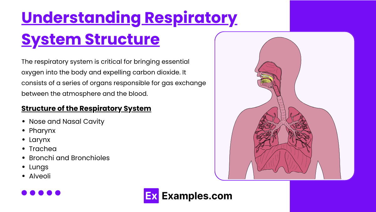
The respiratory system is a network of organs and tissues responsible for gas exchange between the body and the environment. Its primary function is to deliver oxygen to the blood and remove carbon dioxide. The system includes the nasal cavity, pharynx, larynx, trachea, bronchi, and lungs. Within the lungs, tiny air sacs called alveoli facilitate the exchange of gases with the bloodstream. The respiratory system also helps regulate blood pH, filters inhaled particles, and protects the airways with mucus and cilia. It plays a vital role in maintaining cellular respiration and overall metabolic processes. Here’s an overview of its structure:
Structure of the Respiratory System
Nose and Nasal Cavity:
The main external opening for air, the nose is lined with hair and mucus to filter dust and pathogens. The nasal cavity also warms and moistens incoming air.
Pharynx:
A muscular tube that serves both respiratory and digestive systems, connecting the nasal cavity and mouth to the larynx and esophagus. It helps in the process of swallowing and provides a pathway for the movement of air.
Larynx:
Located at the entrance to the trachea and functions in voice production. The larynx contains the vocal cords and works as a valve that closes off the trachea during swallowing, preventing food from entering the airway.
Trachea:
A rigid tube leading from the larynx to the bronchi, it provides a clear airway for air to enter and exit the lungs. The trachea is lined with cilia and mucus to trap and expel contaminants.
Bronchi and Bronchioles:
The trachea divides into two primary bronchi, one entering each lung. These bronchi branch into smaller bronchioles within the lung tissue, further dividing into even smaller passages that lead to the alveoli.
Lungs:
Two large respiratory organs located in the thorax. They house the bronchi, bronchioles, and alveoli. The right lung is typically larger and divided into three lobes, while the left lung has two lobes.
Alveoli:
Tiny sac-like structures at the end of the bronchioles where gas exchange occurs. Each alveolus is surrounded by a network of capillaries where oxygen enters the blood and carbon dioxide is removed.
Function of the Respiratory System
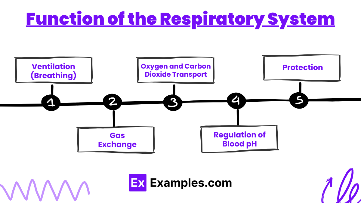
Ventilation (Breathing):
Involves the movement of air in and out of the lungs through the act of inhalation and exhalation. This process is driven by the diaphragm and the intercostal muscles.
Gas Exchange:
Occurs in the alveoli. Oxygen from inhaled air diffuses into the capillaries, while carbon dioxide from the blood diffuses into the alveoli to be exhaled.
Oxygen and Carbon Dioxide Transport:
Blood transports oxygen from the lungs to the cells of the body and brings carbon dioxide back to the lungs to be exhaled.
Regulation of Blood pH:
The respiratory system helps maintain the acid-base balance of the body by regulating the levels of carbon dioxide in the blood, which influences blood pH.
Protection:
Provides defense against pathogens and particulates through the filtering action of nose hairs, mucus, and cilia. Additionally, coughing and sneezing are reflexes that help clear the airways of irritants.
Pathophysiology of Respiratory Disorders
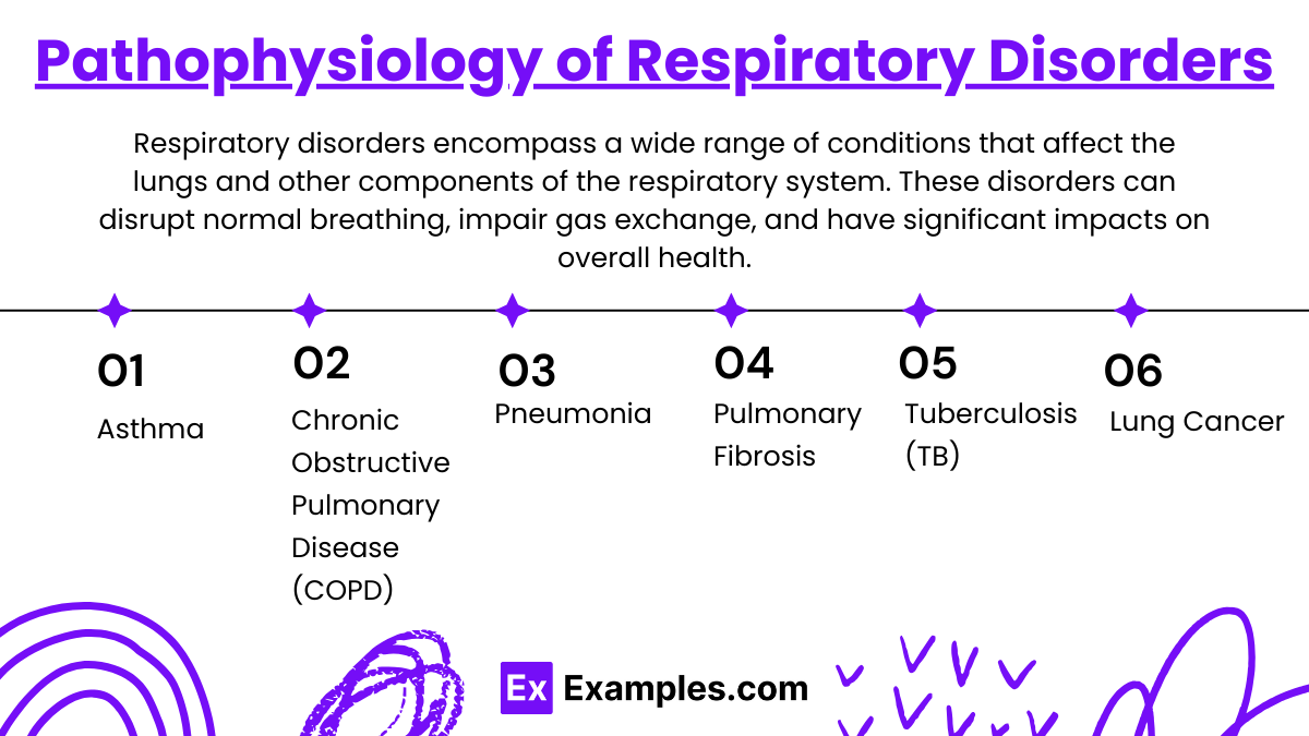
Respiratory disorders encompass a wide range of conditions that affect the lungs and other components of the respiratory system. These disorders can disrupt normal breathing, impair gas exchange, and have significant impacts on overall health. Here's an overview of the pathophysiology behind some common respiratory disorders:
1. Asthma
Pathophysiology: Asthma is characterized by chronic inflammation of the airways, leading to episodes of wheezing, breathlessness, chest tightness, and coughing. During an asthma attack, the airways become hyperresponsive, leading to bronchoconstriction, mucosal edema, and increased mucus production, all of which narrow the airways and obstruct airflow.
2. Chronic Obstructive Pulmonary Disease (COPD)
Pathophysiology: COPD includes conditions such as chronic bronchitis and emphysema, often caused by long-term exposure to irritating gases or particulate matter, most commonly from cigarette smoke. Chronic bronchitis involves inflammation of the lining of the bronchial tubes, leading to persistent cough and mucus production. Emphysema involves damage to the alveoli (air sacs in the lungs), reducing the surface area available for gas exchange and causing air to become trapped in the lungs, which impairs breathing.
3. Pneumonia
Pathophysiology: Pneumonia is an infection that inflames the air sacs in one or both lungs. The air sacs may fill with fluid or pus, causing symptoms such as cough with phlegm or pus, fever, chills, and difficulty breathing. The infection can be bacterial, viral, or fungal.
4. Pulmonary Fibrosis
Pathophysiology: Pulmonary fibrosis involves the progressive scarring of lung tissue, which leads to a continuous decline in lung function. The thickening and stiffening of tissue make it difficult to breathe and reduce oxygen transfer from the lungs to the bloodstream.
5. Tuberculosis (TB)
Pathophysiology: Caused by the bacterium Mycobacterium tuberculosis, TB primarily affects the lungs but can also affect other parts of the body. The infection leads to the formation of granulomas—small nodules of immune cells—in the lungs, which can cause tissue damage and reduce lung function.
6. Lung Cancer
Pathophysiology: Lung cancer originates in the lung tissues, typically in the cells lining the air passages. It is often associated with smoking, but it can also occur in non-smokers. The disease interferes with normal respiratory function primarily through the growth of tumors that block air passages and through the systemic effects of cancer.
Examples
Example 1: Gas Exchange During Exercise
Scenario: Increased oxygen demand during vigorous exercise.
Process: During exercise, the respiratory rate and depth of breathing increase to enhance the oxygen intake and carbon dioxide expulsion. This ensures that more oxygen reaches the muscles and more CO2 is removed from the body, meeting the increased metabolic demands.
Example 2: Asthma Attack
Scenario: Exposure to allergens leading to an asthma attack.
Process: In asthma, exposure to specific allergens can trigger an inflammatory response that leads to bronchoconstriction, airway edema, and increased mucus production. This results in difficulty breathing, wheezing, and coughing. Treatment typically involves bronchodilators which relax the airway muscles.
Example 3: COPD and Airflow Limitation
Scenario: Chronic exposure to cigarette smoke leading to COPD.
Process: In COPD, prolonged exposure to irritants such as cigarette smoke causes chronic inflammation in the airways, destruction of alveolar walls, and narrowing of the airways, which impairs airflow both in and out of the lungs, leading to shortness of breath, especially during physical exertion.
Example 4: Carbon Monoxide Poisoning
Scenario: Inhalation of carbon monoxide (CO) in a closed garage.
Process: CO competes with oxygen for binding sites on hemoglobin, forming carboxyhemoglobin, which reduces the blood's oxygen-carrying capacity. This leads to tissue hypoxia and can result in symptoms ranging from headaches and dizziness to death, depending on the exposure level.
Example 5: Acute Respiratory Acidosis
Scenario: Respiratory failure due to severe pneumonia.
Process: Pneumonia can impair gas exchange by filling the alveoli with fluid or pus, reducing the lung’s ability to oxygenate blood and remove carbon dioxide. This buildup of CO2 leads to respiratory acidosis, where the blood becomes too acidic, potentially requiring mechanical ventilation to correct.
Practice Questions
Question 1: Mechanism of Breathing
What primarily drives the inhalation process during normal quiet breathing?
A) Contraction of the abdominal muscles
B) Relaxation of the diaphragm
C) Contraction of the diaphragm
D) Contraction of the pectoral muscles
Correct Answer: C) Contraction of the diaphragm
Explanation:
During normal quiet breathing, inhalation is primarily driven by the contraction of the diaphragm. When the diaphragm contracts, it moves downward, increasing the volume of the thoracic cavity and reducing the pressure inside the lungs relative to the atmosphere, causing air to flow into the lungs.
Question 2: Gas Exchange
Which gas has the highest partial pressure in the alveoli of the lungs?
A) Oxygen (O2)
B) Carbon dioxide (CO2)
C) Nitrogen (N2)
D) Water vapor (H2O)
Correct Answer: A) Oxygen (O2)
Explanation:
In the alveoli of the lungs, oxygen (O2) typically has the highest partial pressure compared to other gases present in inspired air. This high partial pressure gradient between the alveoli and the blood drives the diffusion of oxygen into the blood, facilitating its transport to tissues throughout the body.
Question 3: Respiratory Adjustments in Exercise
During vigorous exercise, what adjustment does the respiratory system primarily make to meet increased oxygen demand?
A) Decrease in respiratory rate
B) Increase in tidal volume and respiratory rate
C) Increase in carbon dioxide output only
D) Decrease in tidal volume
Correct Answer: B) Increase in tidal volume and respiratory rate
Explanation:
During vigorous exercise, the respiratory system adjusts by increasing both tidal volume (the amount of air inhaled and exhaled in a single breath) and respiratory rate (the number of breaths taken per minute). This adjustment maximizes the amount of air exchanged during breathing and improves the body’s ability to increase oxygen intake and carbon dioxide expulsion to meet the elevated metabolic demands.