)
Sight (vision)

Preparing for the MCAT necessitates a profound comprehension of sight (vision), essential for mastering Organ Systems. Understanding the anatomy of the eye, phototransduction, and visual pathways equips you with crucial insights into visual perception, vital for excelling on the MCAT and understanding complex neurosensory functions.
Learning Objects
In studying "Sight (Vision)" for the MCAT, you should develop an understanding of the mechanisms of visual perception, including the anatomy and function of the eye's structures such as the cornea, lens, retina, and optic nerve. Explore the roles of photoreceptors (rods and cones), the process of phototransduction, and how light is converted into electrical signals in the retina. Understand the visual pathways from the retina to the visual cortex, including how information from both eyes is integrated to create binocular vision and depth perception. Evaluate how different visual cues such as color, motion, and form are processed by the brain, and how visual field defects can affect perception.
Introduction to Sight (vision)
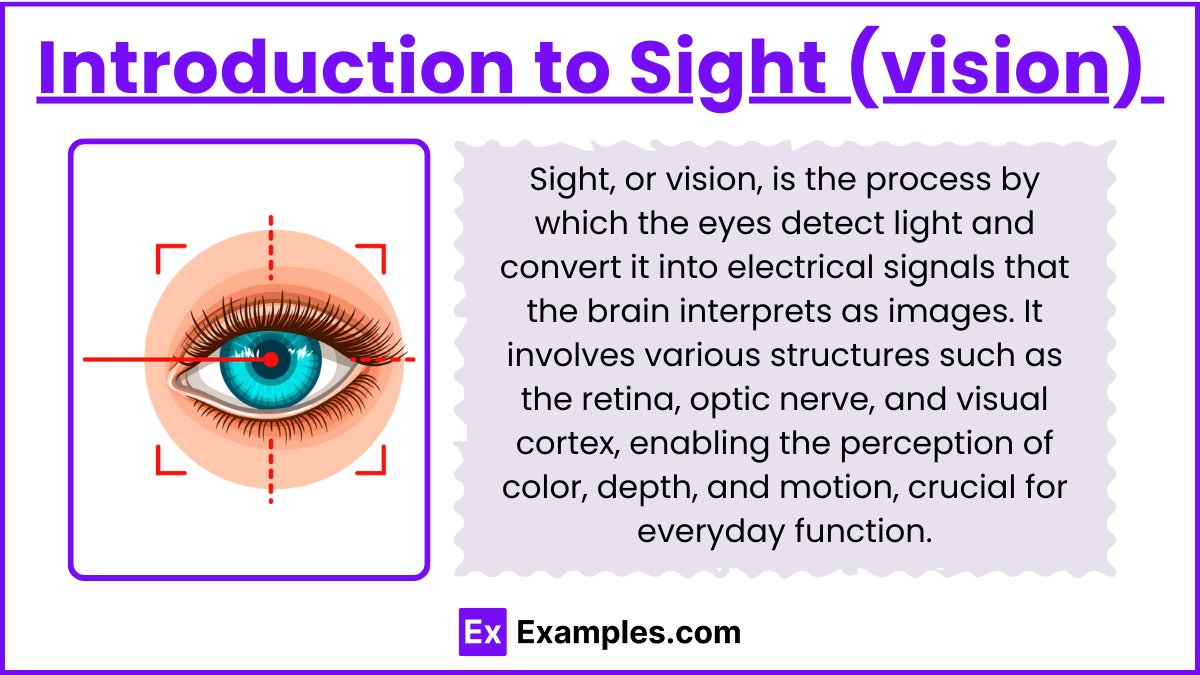
Discover the fundamentals of sight (vision), including eye anatomy, phototransduction, visual pathways, and perception processes, essential for understanding sensory systems on the MCAT.
1. Anatomy of the Eye
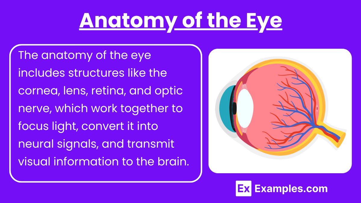
The eye is the primary organ responsible for vision, and its structure is essential for converting light into neural signals.
Cornea: Transparent front layer that refracts (bends) light as it enters the eye.
Pupil: Adjustable opening in the center of the iris that controls the amount of light entering the eye.
Lens: Focuses light onto the retina; changes shape to adjust for near and far vision (accommodation).
Retina: Layer at the back of the eye containing photoreceptor cells (rods and cones) that detect light and convert it into neural signals.
Optic Nerve: Transmits visual information from the retina to the brain.
2. Photoreceptors and Phototransduction
![]()
Photoreceptors in the retina are responsible for detecting light and converting it into electrical signals in a process called phototransduction.
Rods: Photoreceptors sensitive to low light; responsible for night vision and peripheral vision.
Cones: Photoreceptors responsible for color vision and high-acuity vision; three types of cones are sensitive to different wavelengths of light (red, green, blue).
Phototransduction Process:
Light hits photoreceptors (rods and cones), triggering a change in rhodopsin (in rods) or photopsin (in cones).
This change activates a G-protein coupled receptor, transducin, leading to the conversion of cyclic GMP to GMP.
This process leads to the closing of sodium channels, causing hyperpolarization of the photoreceptor.
Hyperpolarization decreases the release of the neurotransmitter glutamate, which in turn modulates the activity of bipolar cells and ganglion cells.
These signals are then transmitted through the optic nerve to the brain.
3. Visual Pathways
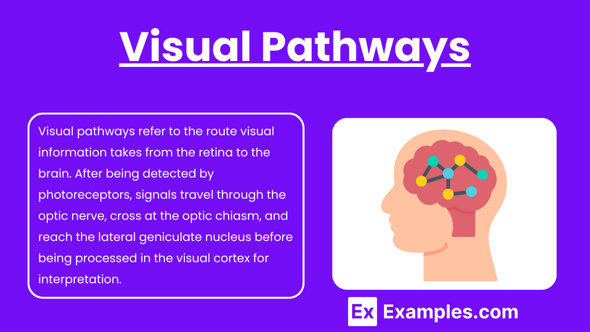
Once light is detected by the retina, the visual information travels through the following pathways:
Optic Nerve: Each eye has an optic nerve that carries signals from the retina to the brain.
Optic Chiasm: The optic nerves from both eyes meet at this junction. Signals from the nasal (inner) side of each retina cross to the opposite side of the brain, while signals from the temporal (outer) side stay on the same side.
Optic Tract: After the optic chiasm, the visual information is carried to the lateral geniculate nucleus (LGN) in the thalamus.
Lateral Geniculate Nucleus (LGN): The LGN processes and relays visual information to the primary visual cortex.
Primary Visual Cortex (V1): Located in the occipital lobe, this region of the brain interprets visual stimuli, including color, form, and motion.
4. Processing Visual Information: Perception and Interpretation
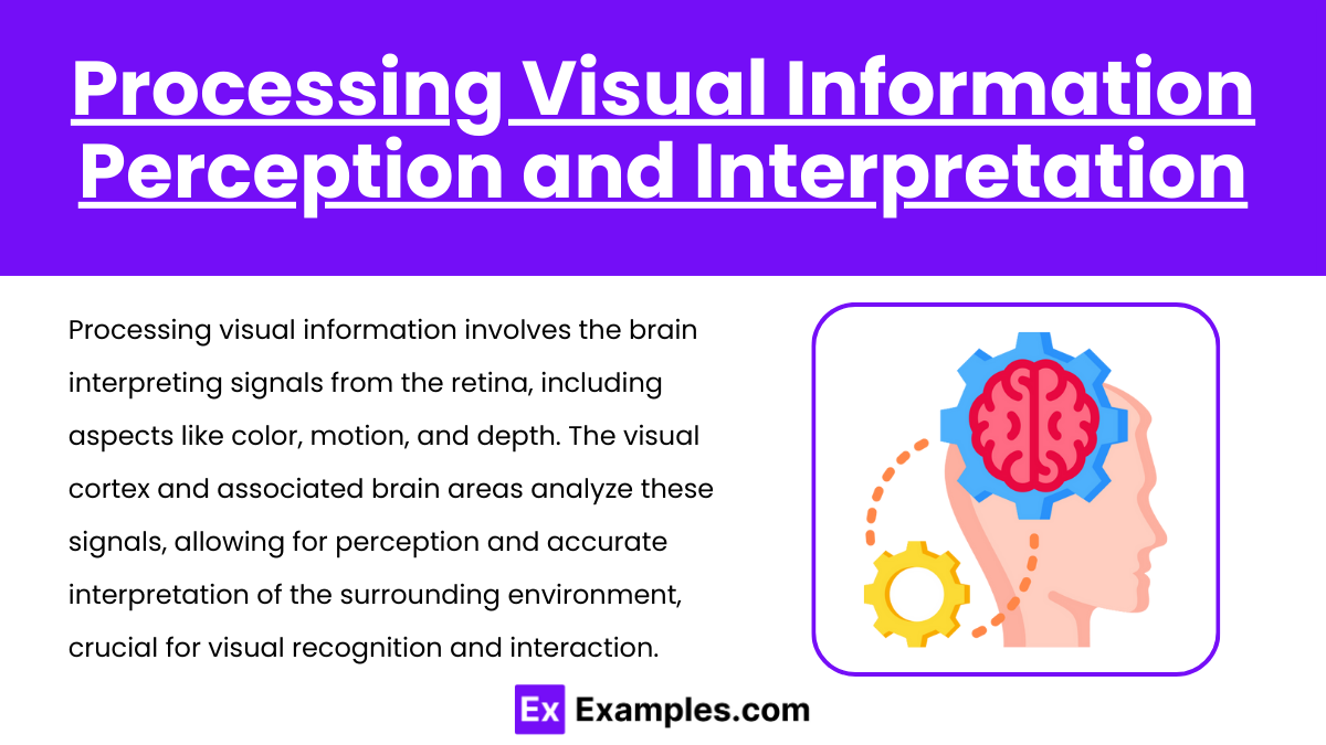
The visual cortex and other associated areas of the brain process the information received from the retina, allowing for detailed perception.
Color Vision: The brain interprets signals from cones to perceive different colors.
Depth Perception: Binocular vision (both eyes) is important for perceiving depth and distance.
Motion Perception: Specialized cells in the retina and brain (such as magnocellular cells) detect motion.
5. Common Disorders Affecting Vision
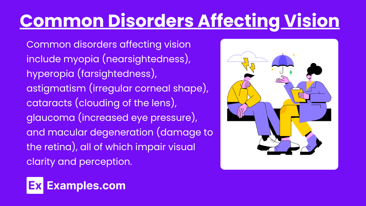
Understanding disorders related to vision is crucial for interpreting MCAT questions on neurosensory dysfunctions.
Myopia (Nearsightedness): Light focuses in front of the retina, causing difficulty in seeing distant objects.
Hyperopia (Farsightedness): Light focuses behind the retina, causing difficulty in seeing close objects.
Astigmatism: Irregular curvature of the cornea or lens, causing distorted vision.
Cataracts: Clouding of the lens, leading to blurry vision.
Glaucoma: Increased pressure in the eye, damaging the optic nerve and leading to vision loss.
Macular Degeneration: Damage to the macula (center of the retina), leading to loss of central vision.
Examples
Example 1: Phototransduction in Low Light
In a dim environment, rods in the retina detect low light levels.
Light hitting the rod cells activates rhodopsin, initiating a biochemical cascade that hyperpolarizes the cell, reducing the release of glutamate.
This process allows the brain to detect visual stimuli in low-light conditions, crucial for night vision.
Example 2: Role of the Optic Nerve in Visual Transmission
The optic nerve carries electrical signals from the retina to the brain.
Information from the nasal half of each retina crosses at the optic chiasm, while information from the temporal halves stays on the same side.
This arrangement is essential for binocular vision and depth perception.
Example 3: Effects of Cataracts on Vision
Cataracts occur when the lens of the eye becomes cloudy, reducing the amount of light reaching the retina.
This leads to blurry vision, difficulty in seeing in bright light, and eventual vision loss if untreated.
Cataract surgery involves replacing the cloudy lens with an artificial one, restoring clear vision.
Example 4: Accommodation for Near Vision
When focusing on nearby objects, the ciliary muscles contract, making the lens more rounded.
This process, known as accommodation, allows light rays to focus properly on the retina for clear near vision.
This ability declines with age, leading to presbyopia, which requires reading glasses for correction.
Example 5: Glaucoma and Increased Intraocular Pressure
Glaucoma results from increased pressure inside the eye due to improper drainage of aqueous humor.
The pressure damages the optic nerve, leading to a gradual loss of peripheral vision.
Early detection and management of intraocular pressure can prevent further vision loss and preserve sight.
Practice Questions
Question 1:
Which of the following correctly describes the role of rods in vision?
A) Detect color vision and function in bright light.
B) Detect motion and are most active during rapid eye movement.
C) Detect low-light conditions and are responsible for night vision.
D) Detect detail and contribute to high-acuity vision.
Answer: C) Detect low-light conditions and are responsible for night vision.
Explanation:
Rods are photoreceptors in the retina that are highly sensitive to low light levels, making them essential for night vision. Unlike cones, which are responsible for color vision and function in bright light, rods do not detect color but are crucial for seeing in dim conditions. They are located primarily in the peripheral regions of the retina and are not involved in detailed or high-acuity vision.
Question 2:
What happens at the optic chiasm?
A) All visual signals are processed for depth perception.
B) Signals from both eyes completely merge into a single pathway.
C) Visual signals from the nasal halves of the retinas cross to the opposite side of the brain.
D) Visual signals from the temporal halves of the retinas cross to the opposite side of the brain.
Answer: C) Visual signals from the nasal halves of the retinas cross to the opposite side of the brain.
Explanation:
At the optic chiasm, the visual signals from the nasal halves of each retina cross over to the opposite side of the brain. This crossover is critical for binocular vision and depth perception, as it allows the brain to combine images from both eyes. Signals from the temporal halves of the retinas remain on the same side, and this arrangement ensures that visual information is processed correctly by the visual cortex for each field of view.
Question 3:
Which process describes the eye's ability to focus on objects at different distances by changing the shape of the lens?
A) Phototransduction
B) Accommodation
C) Refraction
D) Photoreception
Answer: B) Accommodation
Explanation:
Accommodation is the process by which the lens changes its shape to focus light from objects at varying distances onto the retina. When focusing on nearby objects, the ciliary muscles contract, making the lens more rounded for proper focus. This ability is essential for clear vision at different distances and decreases with age, leading to conditions like presbyopia, where reading glasses may be required for close vision. Refraction refers to the bending of light, while phototransduction and photoreception are processes involving light detection by the retina.