Preparing for the MCAT necessitates a profound comprehension of sound (audition), essential for mastering the Nervous and Organ Systems. Understanding the mechanisms of sound waves, frequency, and auditory processing, as well as the role of the cochlea and auditory pathways, equips you with crucial insights into sensory perception. These concepts are vital for excelling on the MCAT and understanding the complex ways in which the brain processes auditory information, crucial for interactions with the environment and communication.
Learning Objective
In studying Sound (Audition) for the MCAT, you should focus on understanding the structure and function of the ear and how sound is transmitted and processed. Analyze how sound waves are converted into electrical signals by the auditory system, and how these signals are interpreted by the brain. Evaluate the role of frequency, amplitude, and pitch in sound perception, as well as the function of structures such as the cochlea and auditory nerve. Additionally, explore how hearing impairments and disorders like tinnitus occur. Apply this knowledge to solve and interpret questions and scenarios in MCAT practice passages, emphasizing the auditory system's role in sensory perception and environmental interaction.
Introduction To Sound (Audition)
Sound (audition) refers to the sensory perception of vibrations or sound waves, which are transmitted through the air and detected by the ear. These waves are converted into electrical signals, allowing the brain to interpret and recognize sounds.
1. Sound Wave Properties
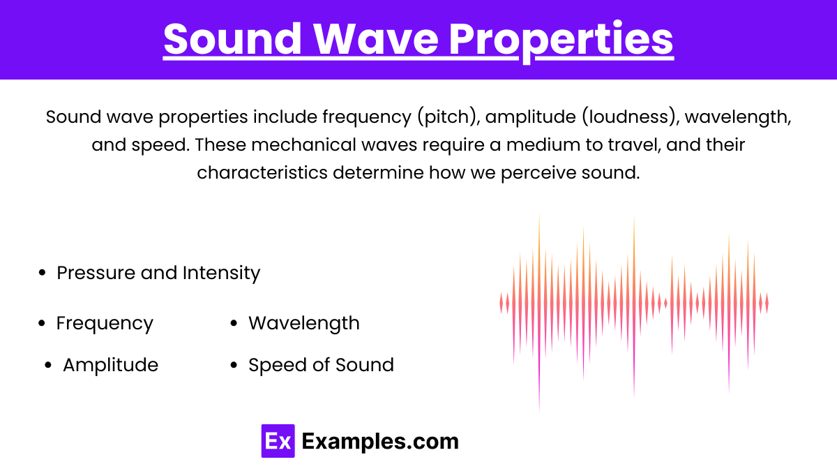
Sound waves are mechanical, longitudinal waves that require a medium (air, liquid, or solid) to propagate. They have several key properties:
Pressure and Intensity: Sound waves involve alternating regions of high pressure (compressions) and low pressure (rarefactions), and intensity is the sound energy transmitted per second per unit area.
Frequency: The number of wave cycles per second, measured in Hertz (Hz), determining the pitch of sound. Higher frequency = higher pitch.
Amplitude: The height of the wave, related to loudness. Larger amplitude means a louder sound, and it’s measured in decibels (dB).
Wavelength: The distance between successive compressions in the wave. Higher frequency = shorter wavelength.
Speed of Sound: Varies based on the medium. It travels faster in solids than in liquids and air. In dry air at room temperature, the speed of sound is approximately 343 m/s.
2. Anatomy of the Ear
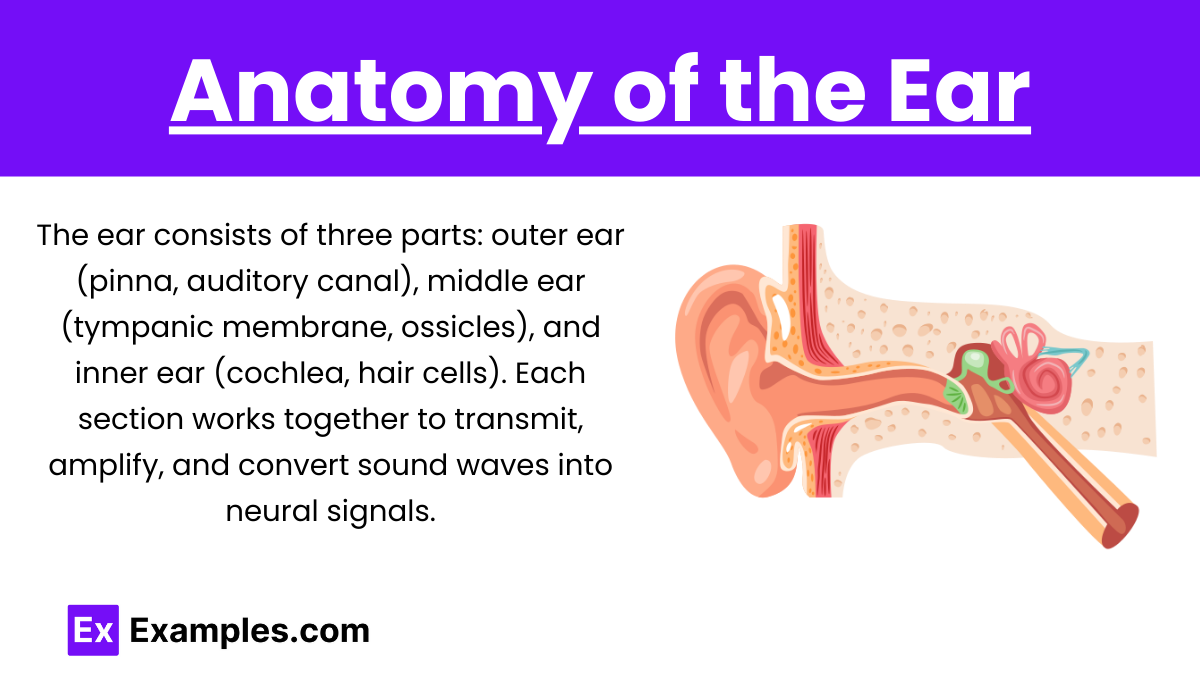
The ear is divided into three main sections that work together to detect and process sound:
Inner Ear: Contains the cochlea, a fluid-filled spiral structure where sound vibrations are converted into electrical signals by hair cells located on the basilar membrane. The inner ear also includes the semicircular canals, which are involved in balance, but not hearing.
Outer Ear: Includes the pinna (auricle) and the external auditory canal. The pinna captures sound waves and directs them into the auditory canal toward the tympanic membrane (eardrum), which vibrates in response to sound.
Middle Ear: Houses the ossicles, three tiny bones called the malleus (hammer), incus (anvil), and stapes (stirrup). These bones amplify the sound vibrations from the eardrum and transfer them to the oval window of the inner ear.
3. Mechanism of Hearing
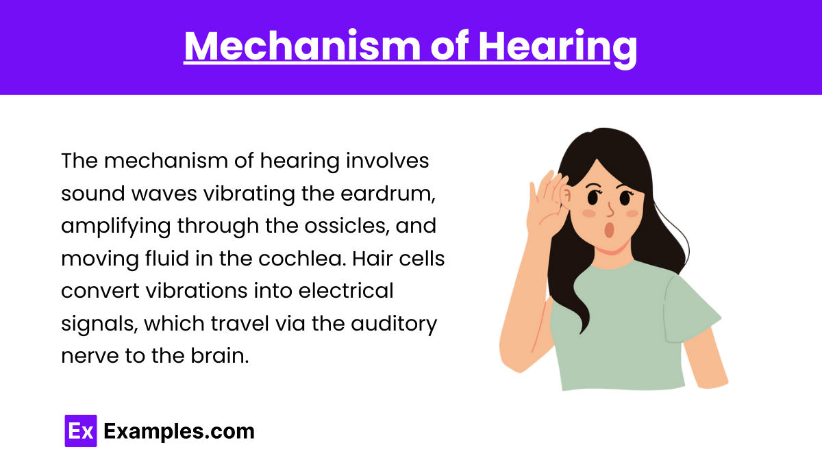
Sound waves enter the ear: The pinna directs sound waves into the auditory canal, causing the tympanic membrane (eardrum) to vibrate.
Vibration amplification: The vibrations from the eardrum are transferred through the ossicles (malleus, incus, stapes), amplifying the sound. The stapes pushes on the oval window of the inner ear.
Cochlear fluid movement: Movement of the oval window causes fluid motion within the cochlea, a spiral-shaped structure in the inner ear. This fluid movement causes the basilar membrane to vibrate.
Hair cell activation: Specialized hair cells on the basilar membrane bend due to the vibrations, converting mechanical energy into electrical signals through the release of neurotransmitters.
Signal transmission: These electrical signals travel through the auditory nerve to the auditory cortex in the brain, where they are processed as recognizable sounds.
4. Auditory Pathways in the Brain
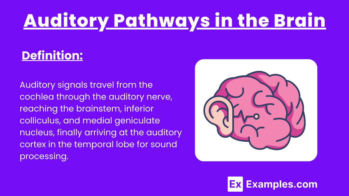
The auditory pathways transmit sound information from the ear to the brain for processing:
Auditory Cortex: Finally, the signals reach the primary auditory cortex in the temporal lobe, where the brain interprets the sounds, recognizing aspects such as pitch, volume, and location.
Cochlear Nerve: Electrical signals generated by the hair cells in the cochlea travel through the cochlear nerve (part of the auditory nerve).
Brainstem (Cochlear Nuclei): The signal reaches the cochlear nuclei in the brainstem, where initial processing and sound localization begin.
Superior Olivary Complex: From the cochlear nuclei, the signals are sent to the superior olivary complex, which helps in binaural hearing by comparing input from both ears to determine the direction of sound.
Inferior Colliculus: Signals are relayed to the inferior colliculus in the midbrain, which integrates auditory information and plays a role in sound reflexes.
Medial Geniculate Nucleus (MGN): The signals then pass through the thalamus, specifically the medial geniculate nucleus, which acts as a relay station, directing the signals to the auditory cortex.
5. Hearing Disorders
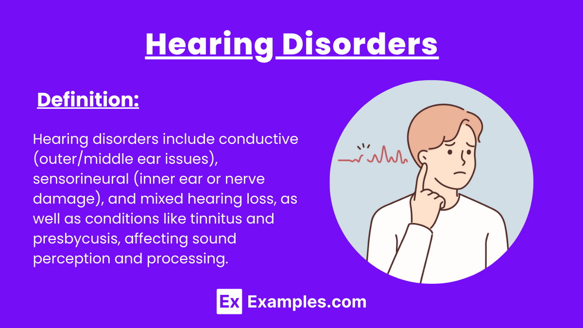
Hearing disorders affect the ability to detect, process, or transmit sound. Common types include:
Conductive Hearing Loss: Occurs when sound is not conducted efficiently through the outer or middle ear. Causes include ear infections, earwax buildup, or damage to the tympanic membrane or ossicles. It affects sound transmission but can often be treated medically or surgically.
Sensorineural Hearing Loss: Results from damage to the inner ear (cochlea) or the auditory nerve. Causes include aging, exposure to loud noises, and genetic factors. Unlike conductive loss, sensorineural hearing loss is usually permanent and managed with hearing aids or cochlear implants.
Mixed Hearing Loss: A combination of both conductive and sensorineural hearing loss, affecting multiple parts of the auditory system.
Tinnitus: A condition characterized by ringing, buzzing, or other phantom sounds in the ears. It’s commonly caused by noise-induced damage to hair cells or other auditory system issues.
Presbycusis: Age-related hearing loss that primarily affects high-frequency sounds due to gradual damage to hair cells in the cochlea.
Examples
Example 1: Sound Transmission in the Outer Ear
Sound waves enter the pinna and are funneled through the auditory canal.
The sound waves cause the tympanic membrane (eardrum) to vibrate, beginning the process of converting sound waves into mechanical vibrations.
Example 2: Vibration Amplification by the Ossicles
Vibrations from the eardrum are transferred to the ossicles (malleus, incus, stapes).
The ossicles amplify these vibrations, passing them to the oval window, which connects to the inner ear.
Example 3: Cochlear Hair Cells and Signal Transduction
Fluid movement in the cochlea causes the basilar membrane to vibrate, bending the hair cells.
Hair cells convert this mechanical movement into electrical impulses, initiating neural signals that travel to the brain.
Example 4: Auditory Pathways to the Brain
Electrical signals travel via the auditory nerve to the brainstem and through the medial geniculate nucleus in the thalamus.
These signals reach the auditory cortex, where they are interpreted as recognizable sounds, including pitch and volume.
Example 5: Impact of Hearing Loss on Auditory Processing
Conductive hearing loss occurs when sound is blocked from reaching the inner ear due to damage to the eardrum or ossicles.
Sensorineural hearing loss involves damage to hair cells in the cochlea, preventing proper signal transmission to the brain.
Practice Test Questions
Question 1:
What structure in the ear is responsible for converting sound vibrations into electrical signals?
A) Tympanic membrane
B) Ossicles
C) Cochlea
D) Pinna
Answer: C) Cochlea
Explanation:
The cochlea in the inner ear contains hair cells that transduce mechanical vibrations into electrical signals. These signals are then transmitted to the brain for processing, allowing us to perceive sound. The tympanic membrane and ossicles are involved in transmitting and amplifying sound waves, but it is the cochlea that performs the critical conversion to electrical signals.
Question 2:
Which of the following best describes the role of the ossicles in hearing?
A) Amplify sound waves before they reach the cochlea
B) Convert sound vibrations into electrical signals
C) Detect sound wave frequencies in the outer ear
D) Protect the inner ear from loud noises
Answer: A) Amplify sound waves before they reach the cochlea
Explanation:
The ossicles (malleus, incus, stapes) are three tiny bones in the middle ear that amplify sound vibrations and transmit them from the tympanic membrane to the oval window of the cochlea. This amplification is crucial for effective hearing, as it enhances the sound signals before they are processed by the inner ear.
Question 3:
What is the primary function of the auditory cortex in the brain?
A) To detect and funnel sound waves into the ear canal
B) To amplify sound waves through the ossicles
C) To process and interpret electrical signals as sound
D) To transduce mechanical energy into neural signals
Answer: C) To process and interpret electrical signals as sound
Explanation:
The auditory cortex, located in the temporal lobe of the brain, is responsible for interpreting the electrical signals received from the ear. It processes the signals, allowing us to recognize different aspects of sound such as pitch, volume, and rhythm.

