Preparing for the NCLEX PN® Exam requires a thorough understanding of the integumentary system, essential for patient care. Mastery of skin anatomy, wound healing, and common conditions such as burns, pressure ulcers, and infections is crucial. This knowledge ensures effective assessment and interventions for maintaining skin integrity and overall health.
Learning Objectives
In studying “Integumentary” for the NCLEX PN® Exam, you should learn to understand the anatomy and physiology of the skin, hair, and nails, including their roles in protection and homeostasis. Analyze common conditions affecting the integumentary system, such as wounds, infections, burns, and pressure ulcers, and recognize their clinical manifestations. Evaluate principles of wound care, infection prevention, and therapeutic interventions. Additionally, explore techniques for assessing skin integrity, identifying risk factors for integumentary disorders, and implementing evidence-based care plans. Apply your understanding to interpreting assessment findings, managing complications, and promoting skin health in diverse patient populations across the lifespan.
Anatomy and Physiology of the Integumentary System

The integumentary system includes the skin, hair, nails, glands, and associated structures, serving as the body’s first line of defense and performing multiple essential functions.
1. Anatomy of the Integumentary System
A. Skin (Primary Organ)
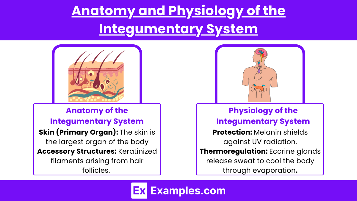
The skin is the largest organ of the body, comprising three main layers:
- Epidermis:
- Outermost layer, made up of keratinized stratified squamous epithelium.
- Avascular, relying on diffusion from the dermis for nutrients.
- Contains specialized cells:
- Keratinocytes: Produce keratin for protection.
- Melanocytes: Produce melanin, which protects against UV radiation.
- Langerhans Cells: Part of the immune system, providing defense against pathogens.
- Merkel Cells: Associated with sensory nerve endings for touch.
- Layers (from superficial to deep):
- Stratum corneum
- Stratum lucidum (in thick skin only)
- Stratum granulosum
- Stratum spinosum
- Stratum basale
- Dermis:
- Middle layer, consisting of connective tissue.
- Divided into two regions:
- Papillary Layer: Thin, areolar connective tissue with capillaries, lymphatics, and sensory neurons.
- Reticular Layer: Thick, dense irregular connective tissue containing collagen and elastin fibers for strength and elasticity.
- Houses hair follicles, sweat glands, sebaceous glands, blood vessels, and sensory receptors.
- Hypodermis (Subcutaneous Layer):
- Not technically part of the skin.
- Made of loose connective tissue and fat (adipose tissue).
- Provides insulation, cushioning, and energy storage.
B. Accessory Structures
- Hair:
- Keratinized filaments arising from hair follicles.
- Functions in protection (e.g., scalp, nostrils) and sensory input.
- Nails: Hard, keratinized structures protecting fingertips and enhancing grip.
- Glands:
- Sebaceous Glands: Produce sebum (oil) to lubricate skin and hair.
- Sweat Glands:
- Eccrine Glands: Regulate body temperature by producing sweat.
- Apocrine Glands: Found in axillary and genital regions, activated during puberty.
- Ceruminous Glands: Produce earwax in the ear canal.
- Mammary Glands: Specialized sweat glands producing milk.
2. Physiology of the Integumentary System
A. Protection
- Acts as a physical barrier against mechanical damage, pathogens, and harmful substances.
- Melanin shields against UV radiation.
B. Thermoregulation
- Maintains body temperature through:
- Sweat Secretion: Eccrine glands release sweat to cool the body through evaporation.
- Vasodilation/Vasoconstriction: Blood vessels in the dermis adjust to dissipate or retain heat.
C. Sensory Reception
- Contains sensory receptors for touch, pressure, pain, and temperature.
- Meissner’s Corpuscles: Detect light touch.
- Pacinian Corpuscles: Detect deep pressure and vibration.
- Free Nerve Endings: Detect pain and temperature.
Common Conditions Affecting the Integumentary System

The integumentary system is susceptible to a wide range of conditions due to its exposure to environmental factors, pathogens, and internal health issues. These conditions may affect the skin, hair, nails, or glands.
1. Infectious Conditions
- Bacterial Infections:
- Impetigo: Highly contagious infection characterized by red sores or blisters that rupture and form golden crusts, commonly caused by Staphylococcus aureus or Streptococcus pyogenes.
- Cellulitis: Deep skin infection resulting in redness, warmth, swelling, and tenderness. Often occurs due to breaks in the skin.
- Viral Infections:
- Herpes Simplex Virus (HSV): Causes cold sores (HSV-1) or genital lesions (HSV-2).
- Varicella-Zoster Virus: Responsible for chickenpox and shingles, presenting as a painful, blistering rash.
- Fungal Infections:
- Tinea (Ringworm): Includes athlete’s foot (tinea pedis), jock itch (tinea cruris), and scalp ringworm (tinea capitis). Characterized by itchy, scaly, circular lesions.
- Candidiasis: Overgrowth of Candida albicans, often in warm, moist areas, causing red, itchy patches.
- Parasitic Infections:
- Scabies: Caused by Sarcoptes scabiei mites burrowing into the skin, leading to intense itching and rash.
- Lice Infestation (Pediculosis): Commonly affects the scalp, causing itching and irritation.
2. Inflammatory and Autoimmune Disorders
- Eczema (Atopic Dermatitis): Chronic, inflammatory condition presenting with red, itchy, and scaly patches. Often triggered by allergens or irritants.
- Psoriasis: Autoimmune disorder causing rapid skin cell turnover, leading to thick, silvery scales and inflamed red patches.
- Seborrheic Dermatitis: A chronic condition causing flaky, scaly skin, often on the scalp, face, and chest.
- Lupus Erythematosus: Systemic autoimmune disease that can cause skin rashes, such as the characteristic butterfly rash on the face.
3. Allergic Reactions
- Contact Dermatitis: Skin reaction caused by contact with allergens or irritants, resulting in redness, itching, and blistering.
- Hives (Urticaria): Raised, itchy welts triggered by allergens, stress, or certain medications.
- Angioedema: Swelling beneath the skin, often accompanying hives, potentially affecting the face, throat, or extremities.
Principles of Wound Care and Infection Prevention

Effective wound care and infection prevention are essential for promoting healing, minimizing complications, and reducing the risk of infection. These principles focus on proper assessment, cleaning, and management of wounds to optimize outcomes.
1. Principles of Wound Care
A. Wound Assessment
- Type of Wound: Identify whether the wound is acute (e.g., surgical, traumatic) or chronic (e.g., pressure ulcers, diabetic ulcers).
- Size and Depth: Measure wound dimensions (length, width, and depth).
- Wound Bed: Evaluate for granulation tissue, slough, or necrosis.
- Exudate: Assess type (serous, purulent, sanguineous), volume, and odor.
- Surrounding Skin: Check for signs of maceration, erythema, or edema.
- Signs of Infection: Look for increased pain, warmth, swelling, redness, or purulent drainage.
B. Wound Cleaning
- Irrigation: Use sterile saline or an appropriate cleansing solution to remove debris and bacteria without damaging healthy tissue.
- Debridement:
- Remove necrotic tissue using mechanical, enzymatic, autolytic, or surgical methods.
- Antiseptics:
- Use cautiously as they may delay healing by damaging healthy tissue.
C. Dressing Selection
- Primary Dressing: Directly contacts the wound bed, promoting healing and moisture balance (e.g., hydrocolloids, alginates).
- Secondary Dressing: Provides additional protection, absorbs exudate, and secures the primary dressing.
- Moisture Management: Maintain a moist environment for optimal healing, but avoid excess moisture that may lead to maceration.
D. Promoting Healing
- Nutritional Support: Encourage adequate protein, vitamins (A and C), and minerals (zinc) intake for tissue repair.
- Offloading Pressure: Use specialized mattresses, cushions, or positioning techniques to prevent pressure injuries.
- Pain Management: Address pain to improve patient comfort and cooperation with wound care.
2. Principles of Infection Prevention in Wound Care
A. Hand Hygiene
- Perform hand hygiene before and after wound care using alcohol-based hand rubs or soap and water.
B. Aseptic Technique
- Use sterile gloves, instruments, and dressings during wound care.
- Minimize contamination by maintaining a clean field.
C. Wound Protection
- Cover wounds to prevent contamination from the environment.
- Educate patients on avoiding unnecessary handling of the wound.
D. Antibiotic Stewardship
- Avoid unnecessary use of systemic antibiotics.
- Use topical antimicrobial agents if the wound shows early signs of infection.
E. Early Detection of Infection
- Recognize symptoms like increased pain, delayed healing, or systemic signs such as fever.
- Obtain wound cultures to identify causative organisms and guide treatment.
Examples
Example 1: Pressure Ulcers
Monitor at-risk clients for skin breakdown, especially over bony prominences. Use interventions like repositioning every 2 hours and applying moisture barriers. Stage ulcers to guide treatment.
Example 2: Burns
Assess burn depth (superficial, partial, full thickness) and calculate total body surface area affected. Prioritize airway, fluid resuscitation, and pain management. Prevent infection with sterile dressing changes.
Example 3: Impetigo
Identify honey-colored crusted lesions typically caused by bacterial infection. Teach clients about hygiene and avoiding lesion scratching. Prescribe topical or oral antibiotics for treatment.
Example 4: Psoriasis
Chronic autoimmune condition with scaly, silvery plaques on elbows, knees, or scalp. Manage with topical steroids, UV therapy, and moisturizers. Educate on triggers like stress and infections.
Example 5: Cellulitis
Localized bacterial skin infection presenting with redness, warmth, and swelling. Treat with prescribed antibiotics and elevate the affected limb. Monitor for systemic symptoms like fever.
Practice Questions
A client with a stage II pressure ulcer on the sacrum is receiving wound care. Which dressing is most appropriate to promote healing?
A. Dry gauze dressing.
B. Hydrocolloid dressing.
C. Transparent film dressing.
D. Wet-to-dry dressing.
Answer:
B. Hydrocolloid dressing.
Explanation: Hydrocolloid dressings maintain a moist environment, promoting wound healing for stage II pressure ulcers. Dry gauze and wet-to-dry dressings are not ideal for this stage, and transparent film dressings are more suitable for stage I ulcers.
Question 2
The nurse is providing care to a client with burns covering 30% of their body. Which assessment finding requires immediate intervention?
A. Urine output of 20 mL/hour.
B. Heart rate of 110 beats per minute.
C. Mild edema around the burn site.
D. Pain rating of 8/10.
Answer:
A. Urine output of 20 mL/hour.
Explanation: A urine output less than 30 mL/hour indicates inadequate kidney perfusion, potentially signaling hypovolemia or shock. This requires immediate intervention. Mild edema, increased heart rate, and pain are expected findings in burn clients.
Question 3
A client with herpes zoster (shingles) reports severe pain and burning in the affected area. Which intervention should the nurse prioritize?
A. Administer antiviral medication as prescribed.
B. Apply a cool compress to the affected area.
C. Teach the client to avoid scratching the lesions.
D. Encourage the client to increase fluid intake.
Answer:
A. Administer antiviral medication as prescribed.
Explanation: Antiviral medication is the priority intervention as it reduces the severity and duration of shingles. Cool compresses and avoiding scratching provide symptomatic relief but are not the priority.


